
Detecting Left Ventricular Systolic Dysfunction on Standard 12-Lead ECG Using a Smartphone-Based Deep Neural Network
Overview
This Powerful Medical-led study developed two smartphone-deployable AI-ECG models (PMcardio LVsense) that detect reduced left ventricular ejection fraction (LVEF) on a standard 12-lead ECG, using ECGs and echocardiograms recorded within 24 hours of each other at a large tertiary care center between 2011 and 2021. In an independent validation set of 25,510 ECG–echocardiogram pairs from unique patients, the LVEF <50% model reached an AUC of 0.952 and the LVEF ≤40% model an AUC of 0.963. Negative predictive values were 99.0% and 99.5%, respectively. In patients whose echocardiogram showed normal LVEF, a discordant AI-ECG reading of reduced LVEF was associated with a 6.8-fold higher risk of developing LV systolic dysfunction over a median follow-up of 7.3 years.
Key findings
- In 25,510 independent validation ECG–echocardiogram pairs, the LVEF <50% model achieved an AUC of 0.952 (95% CI 0.947–0.956), with 89.9% sensitivity and 87.5% specificity.
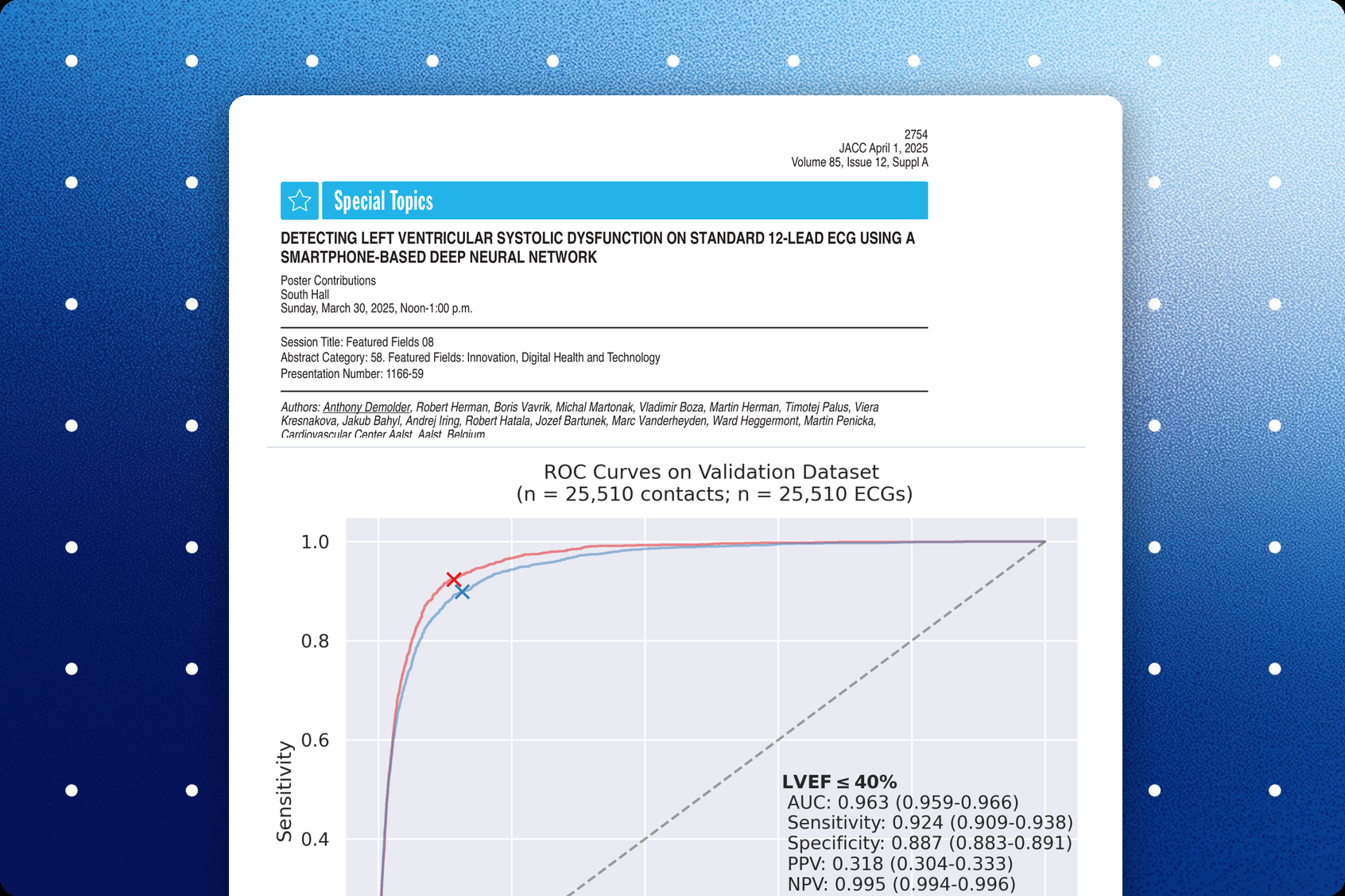
- The LVEF ≤40% model achieved an AUC of 0.963 (95% CI 0.959–0.966), with 92.4% sensitivity and 88.7% specificity.
- Negative predictive values were 99.0% (LVEF <50%) and 99.5% (LVEF ≤40%), with positive predictive values of 38.2% and 31.8%.
- In patients with echocardiography-confirmed normal LVEF, a discordant AI-ECG reading of LVEF <50% was associated with a 6.8-fold increased risk of incident LV systolic dysfunction (HR 6.8, 95% CI 6–7.8; median follow-up 7.3 years).
Published in: ACC.25 – presented as a poster (Journal of the American College of Cardiology, Volume 85, Issue 12, Supplement A)
Published on: 30 March 2025
Background
Point-of-care tools for rapid evaluation of left ventricular (LV) systolic function are lacking. The authors aimed to develop two artificial intelligence (AI) models that detect reduced left ventricular ejection fraction (LVEF) on the 12-lead electrocardiogram (ECG) and that can be deployed using smartphones.
Methods
All ECGs and transthoracic echocardiograms (TTEs) recorded within a 24-hour window between 2011 and 2021 were retrieved from a large tertiary care center. Two independent AI-ECG models were developed to detect LVEF <50% and LVEF ≤40%, and both models were integrated with smartphone-based ECG digitization technology. Key performance metrics included the area under the receiver operating characteristic curve (AUC), sensitivity, specificity, positive predictive value (PPV), negative predictive value (NPV) and F1 score.
Results
A total of 1,205,370 ECGs from 198,274 patients and 291,433 TTEs from 105,849 patients were collected. After pairing ECGs with TTEs, 109,809 ECG–TTE pairs were obtained from 56,236 patients. The independent validation dataset included 25,510 unique ECG–TTE pairs from unique patients. The LVEF <50% model achieved an AUC of 0.952 (95% CI 0.947–0.956), with sensitivity 0.899 (95% CI 0.886–0.912), specificity 0.875 (0.871–0.879), PPV 0.382 (0.368–0.395) and NPV 0.990 (0.989–0.992). The LVEF ≤40% model achieved an AUC of 0.963 (95% CI 0.959–0.966), with sensitivity 0.924 (95% CI 0.910–0.937), specificity 0.887 (0.883–0.891), PPV 0.318 (0.304–0.333) and NPV 0.995 (0.994–0.996). In individuals with echocardiography-confirmed normal LVEF (>50%), a discordant AI-ECG reading of reduced LVEF (<50%) was associated with a 6.8-fold increased risk of developing incident LV systolic dysfunction (HR 6.8, 95% CI 6–7.8; median follow-up 7.3 years).
Conclusion
Both AI models effectively detect reduced LVEF on 12-lead ECGs and are readily accessible via smartphone, providing a rapid and accessible screening tool. This approach enables widescale assessment of LV function in a non-specialist environment and facilitates early identification of patients who may benefit from echocardiographic evaluation.
Share this article



About PMcardio
PMcardio is the market leader in AI-powered diagnostics, addressing the world’s leading cause of death – cardiovascular diseases. The innovative clinical assistant empowers healthcare professionals to detect up to 40 cardiovascular diseases. In the form of a smartphone application, the certified Class IIb medical device interprets any 12-lead ECG image in under 5 seconds to provide accurate diagnoses and individualized treatment recommendations tailored to each patient.

About Powerful Medical
Powerful Medical leads one of the most important shifts in modern medicine by augmenting human-made clinical decisions with artificial intelligence. Our primary focus is on cardiovascular diseases, the world’s leading cause of death.
Established in 2017, Powerful Medical has embarked on a mission to revolutionize the diagnosis and treatment of cardiovascular diseases. We are a medical company backed by 28 world-class cardiologists and led by our expert Scientific Board with decades of experience in daily patient care, clinical research, and medical devices. The results of our research are implemented, developed, certified, and brought to market by our 50+ strong interdisciplinary team of physicians, data scientists, AI experts, software engineers, regulatory specialists, and commercial teams.