

Detect reduced LVEF on ECG with LVsense™ AI ECG.
Identify left ventricular systolic dysfunction (LVEF≤40%) in seconds, directly from a standard 12-lead ECG. Validated on 25,000+ ECG–echo pairs.
- Try for FREE
in the PMcardio App! 

LVsense™ is considered an investigational device in the US.
Not for clinical use.

- 0.963Area under the curve (AUC)
- 92.4%Sensitivity
- 88.7%Specificity
Performance metrics from internal validation studies.
LVsense™ is a clinically validated AI ECG model that detects reduced left ventricular ejection fraction (LVEF≤40%) directly from a standard 12-lead ECG. It gives clinicians a fast, non-invasive signal of impaired heart function at first patient contact, helping identify who needs confirmatory echocardiography.
Heart failure is common, consequential, and routinely caught too late
Accurate and timely assessment of left ventricular systolic function, measured by LVEF, is essential for optimal management and better patient outcomes.¹ Echocardiography measures it accurately but isn't available at most points of first contact, and early disease is frequently silent. The result is a large population with treatable, reduced LVEF identified only after symptoms, hospitalization, or deterioration.
- 3.4%Up to 3.4% of the population have asymptomatic reduced LV systolic function³
- 2%2% of adults live with heart failure, rising above 10% in those aged 70+⁵
- 53–67%5-year mortality once heart failure is established⁶
- —Limited echocardiography access leaves reduced LVEF undetected¹
LVsense™ AI detects reduced LVEF with 92.4% sensitivity and 88.7% specificity, directly from a 12-lead ECG
A negative LVsense result carries a 99.5% negative predictive value, supporting rule-out of reduced LVEF at the prevalence studied.
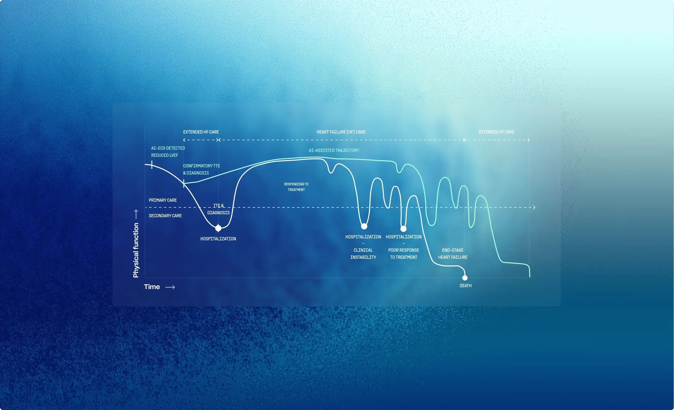
The role of patient screening — the AI-ECG patient-trajectory diagram vs standard care.
Routine screening using the AI-augmented 12-lead ECG allows for swift assessment of LV function, helping accurately identify patients needing confirmatory echocardiography.⁷
A rapid, non-invasive read on heart function, built on deep learning and echo-confirmed labels.
Beyond the human eye
LVsense detects patterns of reduced LV systolic function that are not visible to clinicians on a standard ECG, a capability beyond many physicians' skills.
Echo-confirmed reference standard
Training and validation labels were derived from transthoracic echocardiography, using ECG–echo pairs within a 24-hour interval to maximize the correspondence between electrical and cardiac functional status.
Trained at scale
Developed on a data vault of more than 1.2 million ECGs and ~300,000 echocardiograms, yielding ~100,000 paired ECG–echo records across >50,000 unique patients.⁷
Fixed Model
LVsense is a fixed, locked algorithm that does not change or retrain in clinical use, providing stable and reproducible predictions without generating hallucinated outputs.
Capture
Clinician photographs a standard 12-lead ECG in the PMcardio app.

Analyse
LVsense digitizes the ECG and evaluates LV systolic function. App returns a reduced-LVEF signal with a confidence score; informative ECG regions highlighted.

Act
Patient prioritized for confirmatory echo and earlier specialist referral.
Impact of LVsense™
Clinical and operational benefits
- Earlier heart failure diagnosis
- Surfaces impaired LV function from an ECG taken for any reason — catching heart failure before it forces its way into the ED.²
- Guideline-directed medical therapy, sooner
- Gets reduced-LVEF patients onto guideline-directed medical therapy (GDMT) at the point it changes their trajectory most.³
- Reduced HF hospitalizations
- Early diagnosis and adequate treatment reduce acute exacerbations and complications of HF, minimizing heart failure hospitalizations.³
- Cost savings
- Minimizes preventable hospitalizations, reducing overall healthcare expenditures.
- A screening pathway that scales
- By identifying reduced LV systolic function early, an improved patient pathway can be created. LVsense™ puts a heart-function screen at every first point of contact — without adding echo machines or sonographers.
PMcardio combines clinically validated AI diagnostics with workflow-focused tools, extending reduced-LVEF detection from the echo lab to the first point of contact.
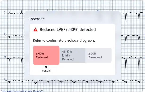
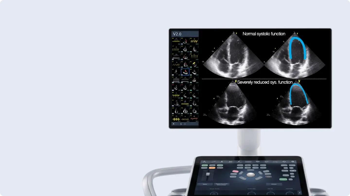
AI-Driven LVEF Assessment
LVsense™ assesses LV systolic function directly from a 12-lead ECG, a capability beyond clinicians' ECG reading.
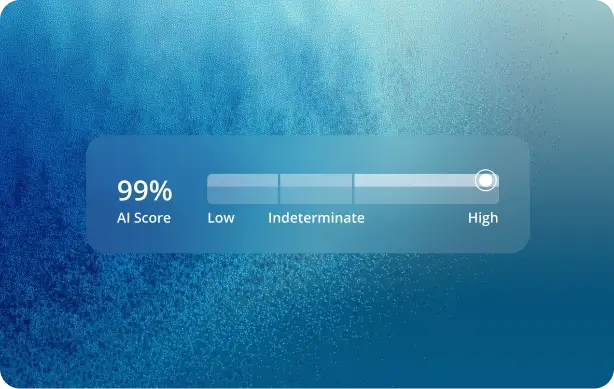
AI Confidence Score
Provides a probability-based confidence score to support clinical decision-making in borderline or ambiguous ECG cases.

Reliable Rule-Out for Screening
High negative predictive value supports rule-out of reduced LVEF in the screened population, focusing echo capacity on higher-probability patients.

Faster Time to Assessment
Collapses the time required to assess LV function, enabling faster clinical decision-making and timely referral for confirmatory echo.
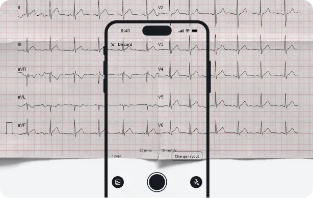
Works With Any 12-Lead ECG Image
Coupled with PMcardio's smartphone ECG digitization, LVsense works from a photo of a printed or on-screen 12-lead ECG and is compatible with existing ECG devices & workflows.
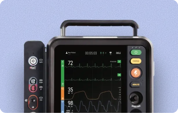
Environment-agnostic
Designed to analyze standard 12-lead ECGs from a wide range of ECG devices used in primary, emergency, and hospital settings.
Plug LVsense™ in across the
hub-and-spoke network
AI-ECG screening for reduced LVEF from a 12-lead ECG — at every point of care, routed to confirmatory echo at the hub

Validated Against Echocardiography in a Large, Real-World Dataset
LVsense™ was developed and validated on echo-paired 12-lead ECGs and presented at major international cardiology congresses.⁷
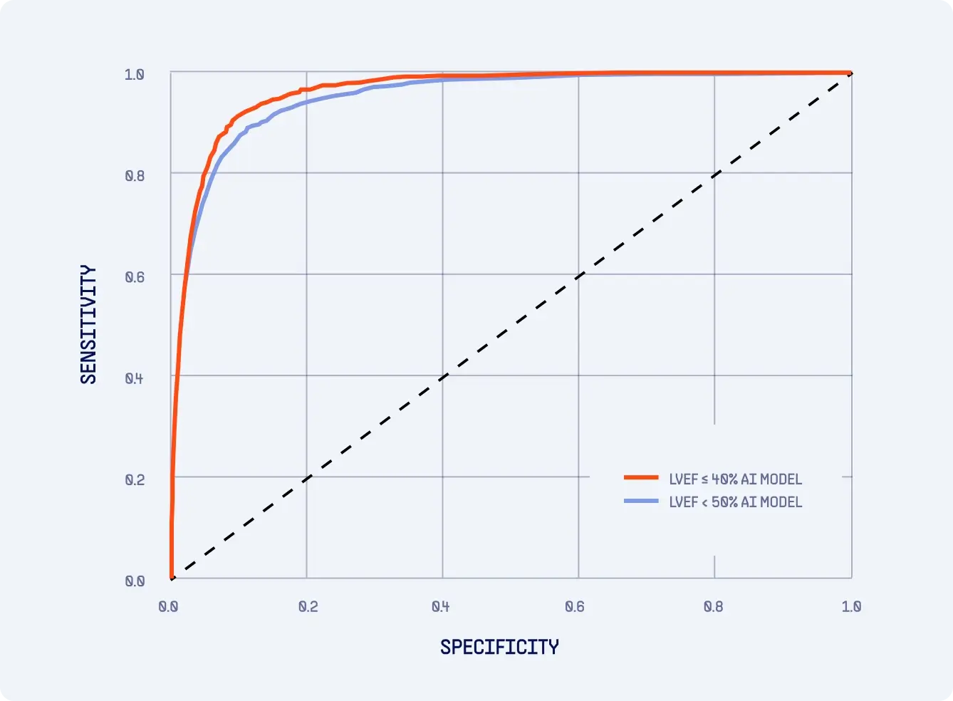
Headline Validation — LVEF≤40% Model
- 0.963 (95% CI 0.959–0.966) AUC
- 92.4% (95% CI 90.9–93.8) Sensitivity
- 99.5% (95% CI 99.4–99.6) Negative Predictive Value
- Specifity
- 88.7% (95% CI 88.3–89.1)
- PPV
- 31.8% (95% CI 30.4–33.3)
Headline Validation — LVEF<50% Model
- 0.952 (95% CI 0.947–0.956) AUC
- 89.8% Sensitivity
- 99.0% Negative Predictive Value
- Specifity
- 87.5%
Study Parameters
- Design
- retrospective; ECGs paired with transthoracic echocardiograms within a 24-hour window
- Data pooled
- >1,2 milion ECGs and >300.000 TTEs → >100.000 ECG–TTE pairs across >60.000 unique patients
- Validation set
- >25.000 distinct TTE–ECG pairs (>25.000 patients)
- Prevalence (validation set)
- lVEF ≤ 40% = 5.4%
- peference standard
- echocardiographic LVEF
Prospective Studies Evaluating Real-World Impact
LVsense™ is being studied in real-world populations and care pathways to evaluate clinical and operational impact.
Multicenter validation study
Prospective multisite study
Populations
General population screening
Emergency department triage
Cardiotoxicity screening
Pneumology wards
Diabetes clinics

LVsense™ is one of PMcardio's clinically validated AI ECG models and is being adopted across a growing international network of hospitals, bringing point-of-care reduced-LVEF screening closer to first patient contact.
Join more than 30 hospitals leveraging AI to streamline cardiac care and improve patient outcomes.

Reimbursement pathway for AI-ECG
AI-based ECG analysis now has a defined coding and payment route. In the US, the AMA introduced Category III CPT codes 0764T and 0765T for AI-powered ECG analysis to detect cardiac pathology, and from January 1, 2025, CMS set a national hospital-outpatient payment for them (APC 5734) — the first national reimbursement for AI-ECG detection of conditions such as reduced ejection fraction. The 2026 CPT set adds further codes for algorithmic ECG detection of cardiac dysfunction. Together, they give hospitals a clearer financial path to adopt AI-ECG at the point of care.
Reimbursement & Coding
- CODE
- DESCRIPTION
- 0764T
- AI-ECG algorithmic assessment for cardiac dysfunction (e.g., low EF); concurrently performed ECG
- 0765T
- AI-ECG algorithmic assessment for cardiac dysfunction (e.g., low EF); concurrently performed ECG
- 0765T
- AI-ECG algorithmic assessment for cardiac dysfunction; using a previously performed ECG
- 93000
- Electrocardiogram, routine ECG with ≥12 leads; with interpretation and report
- 93010
- Electrocardiogram, 12 leads; interpretation and report only
0764T / 0765T are Category III (emerging-technology) codes; in the US, CMS set a national hospital-outpatient payment (APC 5734) for them, effective Jan 1, 2025. Codes and coverage vary by payer and setting — check the CMS Physician Fee Schedule. LVsense™ is an investigational device in the US.
Frequently Asked Questions
What does LVsense™ measure and what does "reduced LVEF" mean?
LVsense™ estimates left ventricular systolic function — how effectively the heart's main pumping chamber contracts — directly from a 12-lead ECG. It flags reduced LVEF, defined as a left ventricular ejection fraction ≤ 40%. Ejection fraction is the share of blood the left ventricle pumps out with each beat; a normal value is roughly ≥ 50%, so ≤ 40% indicates impaired pumping (left ventricular systolic dysfunction), the hallmark of heart failure with reduced ejection fraction. Detecting it earlier helps route patients to timely confirmation and treatment.
What is a normal ejection fraction and what counts as reduced?
A normal left ventricular ejection fraction (LVEF) is generally 50–70%. An LVEF of 41–49% is considered mildly reduced, and 40% or below is classified as reduced. LVsense™ is built to flag the patients most at risk — those with an LVEF of 40% or lower.
What are the symptoms of reduced ejection fraction?
Common symptoms include fatigue, breathlessness on exertion, and swelling in the legs or ankles. The difficulty is that these signs are nonspecific and often absent in early disease — a meaningful share of people with reduced LVEF have no symptoms at the stage where treatment could most change their outcome. That silent window is exactly why a point-of-care screen matters.
Does LVsense™ replace echocardiography?
No. Echocardiography remains the reference standard for measuring LVEF. LVsense™ is a screening signal, not a diagnosis — it helps identify which patients should be prioritized for confirmatory echo. Because a negative result carries a high negative predictive value (99.5% at the prevalence studied), it supports rule-out and helps focus limited echo capacity on higher-probability patients. A positive flag should always be confirmed by echocardiography.
What ECG input does LVsense™ need?
A standard resting 12-lead ECG. No proprietary hardware is required — LVsense™ works from a smartphone photo of a printed or on-screen ECG, which the PMcardio app digitizes before analysis. It is designed to work with ECGs from the wide range of standard devices used in primary care, emergency, and hospital settings. For health systems, PMcardio also integrates with existing clinical infrastructure, so ECGs and results can flow directly to and from the electronic health record (EHR) without disrupting current workflows.
How accurate is LVsense™?
Validated against echocardiography (LVEF ≤ 40% model), LVsense™ achieved an AUC of 0.963, sensitivity of 92.4%, specificity of 88.7%, and a negative predictive value of 99.5%, evaluated on 25,510 echo-paired ECGs. Full methods, the secondary LVEF < 50% model, and peer-reviewed results are available on our publications page.
Is LVsense™ available in my region / authorized by the FDA?
Availability depends on your region. In the EU and UK, LVsense™ is provided as part of PMcardio's CE-marked AI-ECG modules under the EU Medical Device Regulation (MDR). In the United States, the LVEF AI ECG Model (LVsense™) is considered an investigational device and is not for clinical use. US authorization to date covers the Queen of Hearts™ (ACS AI ECG Module), which has received De Novo authorization from the U.S. Food and Drug Administration (FDA); it does not extend to LVsense™. Not all modules of the PMcardio platform are available in every region.
How do I get access and what does it cost?
LVsense™ is available in the PMcardio app — download it from the App Store or Google Play and try it free, with 5 free ECGs per month and no credit card required. Plans for individuals and organisations are listed on the Pricing page; to roll it out across a hospital or network, use Get access to start.
Be at the Forefront of Cardiac Care. Join the AI-driven LVEF Detection Revolution Today!
References
- Lang RM, Badano LP, Mor-Avi V, et al. Recommendations for cardiac chamber quantification by echocardiography in adults: an update from the American Society of Echocardiography and the European Association of Cardiovascular Imaging. Eur Heart J Cardiovasc Imaging. 2015;16(3):233-70.
- Marwick TH, Gillebert TC, Aurigemma G, et al. Recommendations on the use of echocardiography in adult hypertension: a report from the EACVI and the ASE. Eur Heart J Cardiovasc Imaging. 2015;16(6):577-605.
- SOLVD Investigators, Yusuf S, Pitt B, Davis CE, Hood WB Jr, Cohn JN. Effect of enalapril on mortality and the development of heart failure in asymptomatic patients with reduced left ventricular ejection fractions. N Engl J Med. 1992;327(10):685-91.
- Ledwidge M, Gallagher J, Conlon C, et al. Natriuretic peptide–based screening and collaborative care for heart failure: the STOP-HF randomized trial. JAMA. 2013;310(1):66-74.
- Dunlay SM, Roger VL, Redfield MM. Epidemiology of heart failure with preserved ejection fraction. Nat Rev Cardiol. 2017;14(10):591-602.
- Benjamin EJ, Levy D, Vasan RS, et al. Impact of heart failure on mortality rates in the Framingham Heart Study population: an update. JAMA. 1999;281(18):1728-34.
- Demolder A, Herman R, Vavrik B, Martonak M, Boza V, Herman M, Palus T, Kresnakova V, Bahyl J, Iring A, Hatala R, Bartunek J, Vanderheyden M, Heggermont W, Penicka M. A smartphone-based AI model to detect left ventricular systolic dysfunction on 12-lead ECG. Abstract. Eur Heart J. In press.

Queen of Hearts
AI-powered detection of STEMI and occlusive MI — including subtle STEMI equivalents that standard criteria miss.

Core AI
Comprehensive ECG analysis — rhythms, conduction abnormalities, blocks, hypertrophies, and interval measurements.