
A smartphone based AI model to detect left ventricular systolic dysfunction on 12-lead ECG
Overview
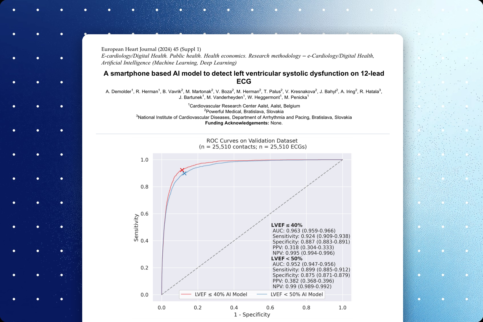
This study from the Cardiovascular Research Center Aalst, Belgium, and Powerful Medical developed and validated two AI-ECG models (PMcardio LVsense) that detect reduced left ventricular ejection fraction (LVEF) on a single 12-lead ECG captured with a smartphone. ECGs and echocardiograms recorded within 24 hours of each other between 2011 and 2021 were randomly split into development and validation sets, with 25,510 patients in the validation set. The LVEF ≤40% model reached an AUC of 0.963, with 92.4% sensitivity, 88.7% specificity and a negative predictive value of 99.5%, and the LVEF <50% model reached an AUC of 0.952. The authors see the models as a preliminary screening tool to select patients for confirmatory echocardiography.
Key findings
- In 25,510 validation patients, the LVEF ≤40% model achieved an AUC of 0.963 (95% CI 0.959–0.966), with 92.4% sensitivity and 88.7% specificity.
- The LVEF <50% model achieved an AUC of 0.952 (95% CI 0.947–0.956), with 89.9% sensitivity and 87.5% specificity.
- At a prevalence of 5.4% for LVEF ≤40%, the model's negative predictive value was 99.5% and its positive predictive value 31.8%.
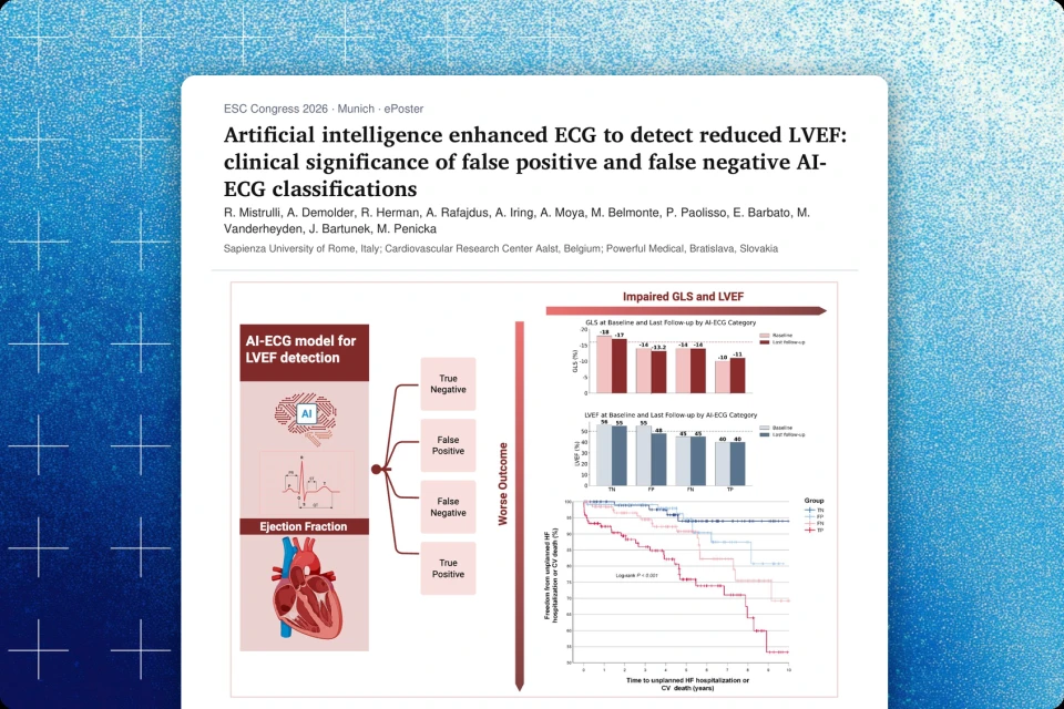
- In the congress presentation, patients with normal LVEF on echocardiography whom the AI flagged as having reduced LVEF had a hazard ratio of 6.8 for later developing LVEF <50% over a median follow-up of 7.3 years.
Published in: ESC Congress 2024, London – presented by A. Demolder; abstract published in European Heart Journal, Volume 45, Supplement 1
Published on: 1 September 2024
Background
Although echocardiography is essential for cardiac assessment, current clinical practice lacks a point-of-care tool for rapid evaluation of left ventricular ejection fraction (LVEF). Left ventricular systolic dysfunction is often diagnosed only once it has become symptomatic, even though earlier diagnosis is desirable and effective treatments exist. The authors therefore set out to develop and validate two artificial intelligence (AI) models that accurately detect reduced LVEF on a single 12-lead ECG captured with a smartphone, to help identify patients at risk of heart failure who may benefit from more detailed echocardiographic evaluation.
Methods
A large collection of ECGs and transthoracic echocardiograms (TTEs) recorded between 2011 and 2021 was sourced from the hospital data vault of OLV Hospital Aalst, Belgium. ECGs were paired with TTEs recorded within a 24-hour window, with biplane Simpson LVEF given priority over visually estimated LVEF, and the resulting pairs were randomly assigned to a model development dataset (50%) and a validation dataset (50%); only one ECG-TTE pair per patient was allowed for validation. Two AI-ECG models were developed, one to detect LVEF ≤40% and another to detect LVEF <50%, based on the ESC definitions of heart failure. Both models were paired with smartphone-based ECG digitization technology. Performance was assessed by the area under the receiver operating characteristic curve (AUC), sensitivity, specificity, positive predictive value (PPV), negative predictive value (NPV) and F1-score.
Results
A total of 1,205,370 ECGs (198,274 patients) and 291,433 TTEs (105,849 patients) were collected and paired, resulting in 109,809 ECG-TTE pairs from 56,236 patients. The validation dataset consisted of 25,510 distinct ECG-TTE pairs from 25,510 patients, with a prevalence of 5.4% for LVEF ≤40% and 7.9% for LVEF <50%. The LVEF ≤40% model achieved an AUC of 0.963 (95% CI 0.959–0.966), sensitivity of 0.924 (0.910–0.937), specificity of 0.887 (0.883–0.891), PPV of 0.318 (0.304–0.333), NPV of 0.995 (0.994–0.996) and F1-score of 0.474 (0.457–0.490). The LVEF <50% model achieved an AUC of 0.952 (0.947–0.956), with a slightly lower sensitivity of 0.899 (0.886–0.912), specificity of 0.875 (0.871–0.879), PPV of 0.382 (0.368–0.395), NPV of 0.990 (0.989–0.992) and F1-score of 0.536 (0.521–0.550). In the congress presentation, performance was consistent across sex, age and BMI subgroups, and patients with normal LVEF on echocardiography but an AI prediction of reduced LVEF had a higher incidence of developing LVEF <50% over a median follow-up of 7.3 years (hazard ratio 6.8).
Conclusion
The AI models can identify reduced LVEF from standard 12-lead ECGs captured with a smartphone. The results suggest potential clinical utility as a preliminary screening tool to select patients for confirmatory echocardiography, offering a timely and cost-effective way to identify patients at risk of developing heart failure.
Share this article



About PMcardio
PMcardio is the market leader in AI-powered diagnostics, addressing the world’s leading cause of death – cardiovascular diseases. The innovative clinical assistant empowers healthcare professionals to detect up to 40 cardiovascular diseases. In the form of a smartphone application, the certified Class IIb medical device interprets any 12-lead ECG image in under 5 seconds to provide accurate diagnoses and individualized treatment recommendations tailored to each patient.

About Powerful Medical
Powerful Medical leads one of the most important shifts in modern medicine by augmenting human-made clinical decisions with artificial intelligence. Our primary focus is on cardiovascular diseases, the world’s leading cause of death.
Established in 2017, Powerful Medical has embarked on a mission to revolutionize the diagnosis and treatment of cardiovascular diseases. We are a medical company backed by 28 world-class cardiologists and led by our expert Scientific Board with decades of experience in daily patient care, clinical research, and medical devices. The results of our research are implemented, developed, certified, and brought to market by our 50+ strong interdisciplinary team of physicians, data scientists, AI experts, software engineers, regulatory specialists, and commercial teams.