
From normal ECG to STEMI Equivalent: an evolving Acute Coronary Syndrome detected by AI
Overview
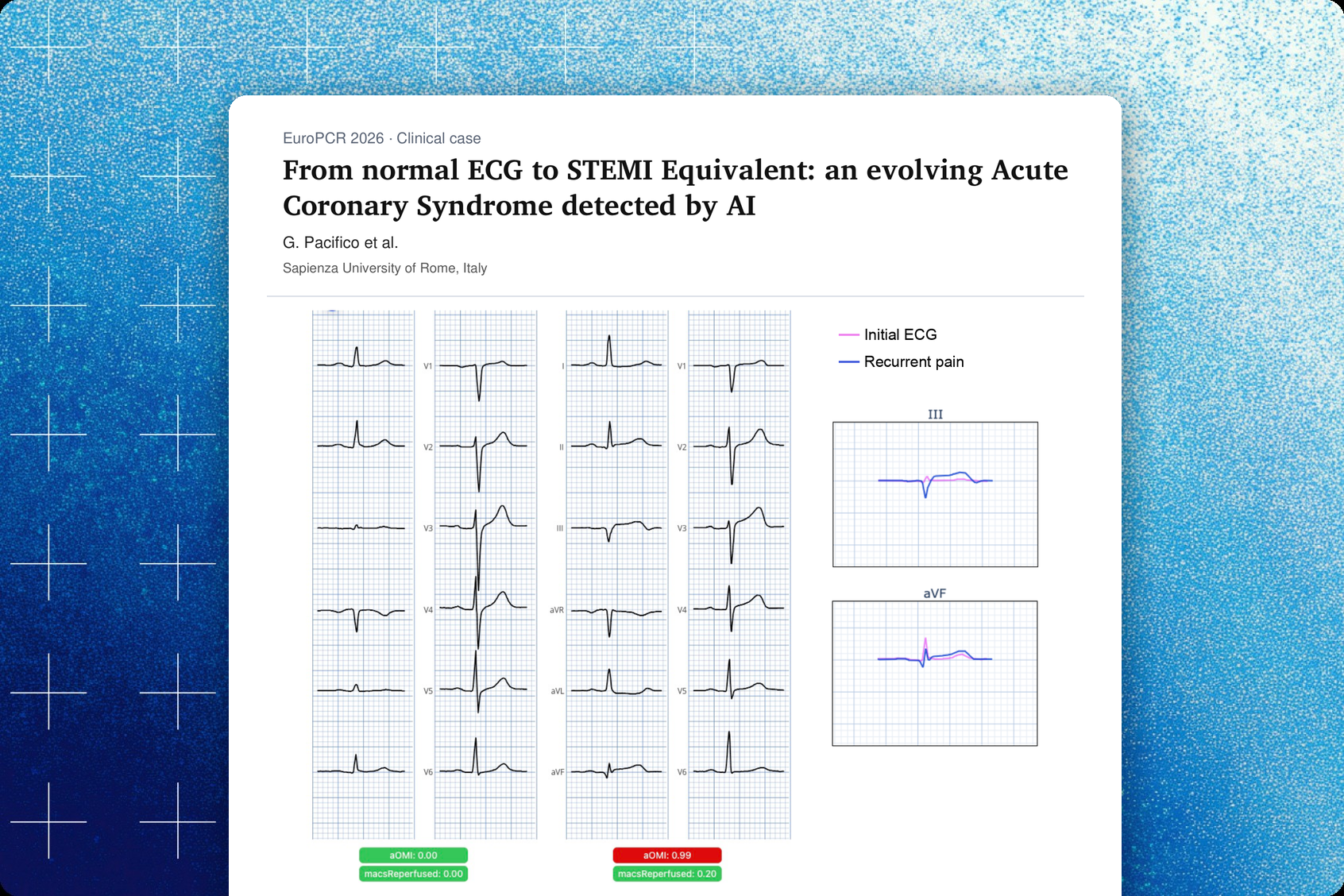
This independent clinical case from Sapienza University of Rome describes a 48-year-old man admitted with oppressive chest pain and dyspnea whose initial ECG, recorded while he was pain-free, appeared normal, and which the PMcardio Queen of Hearts AI-ECG model scored at aOMI 0.00. With a negative high-sensitivity troponin curve and a normal echocardiogram, he underwent coronary CT angiography, which showed complete occlusion of the mid right coronary artery (RCA). When his chest pain recurred, a repeat ECG showed evolving STEMI-equivalent changes and the AI score rose to aOMI 0.99. Primary PCI treated a thrombotic mid-RCA occlusion, followed by staged PCI of the left anterior descending artery (LAD).
Key findings
- The initial ECG, recorded without chest pain, appeared normal and was scored aOMI 0.00 by the Queen of Hearts AI model.
- During recurrent chest pain, the repeat ECG showed STEMI-equivalent changes flagged by the AI model (aOMI 0.99), with high-sensitivity troponin I at 166 pg/mL.
- Coronary CT angiography showed complete occlusion of the mid RCA, and primary PCI treated the thrombotic occlusion with a 2.25 × 22 mm drug-eluting stent plus a 2.25 × 20 mm drug-coated balloon for a residual distal stenosis.
- A moderate proximal LAD stenosis with FFR 0.74 was treated with staged PCI, after which FFR improved to 0.82.
Published in: EuroPCR 2026 – presented as an eCase
Published on: 22 May 2026
Background
STEMI criteria have limited sensitivity for acute coronary occlusion myocardial infarction (OMI), especially when the occlusion is early or intermittent. This case shows how an initially normal ECG can evolve into a STEMI equivalent, and how an AI-ECG model can support the recognition of occlusion on serial ECGs.
Methods
A 48-year-old man with hypertension, class 1 obesity, current smoking and a family history of coronary artery disease, and a history of anaphylactic shock after aspirin, was admitted to the emergency department with oppressive chest pain and dyspnea. His initial ECG, recorded while he was free of chest pain, showed sinus rhythm and appeared normal. The high-sensitivity troponin I curve was negative and transthoracic echocardiography was normal (LVEF 55%, no regional wall motion abnormalities). Given his high-risk profile and typical but resolved angina, coronary CT angiography (CCTA) was performed. Serial ECGs were analyzed with the PMcardio Queen of Hearts AI-ECG model, which reports an aOMI score, and compared side by side.
Results
The AI model scored the initial ECG at aOMI 0.00 and found no STEMI or STEMI-equivalent changes. CCTA showed a patent left main coronary artery, a mixed plaque in the proximal LAD up to the first diagonal branch with moderate stenosis, a proximal calcific plaque with moderate stenosis in the left circumflex artery, and complete occlusion of the RCA from its mid segment. While awaiting further management, the patient developed recurrent chest pain with an evolving ECG; high-sensitivity troponin I was 166 pg/mL. The AI model identified STEMI-equivalent changes on this ECG (aOMI 0.99), and a side-by-side comparison with the first ECG showed the evolution of the changes. Primary PCI of the thrombotic mid-RCA occlusion was performed with a 2.25 × 22 mm drug-eluting stent, and a residual significant distal stenosis was treated with a 2.25 × 20 mm drug-coated balloon, with a good angiographic result. Complete revascularization followed with staged PCI of the proximal LAD: functional assessment showed Pd/Pa 0.91, RFR 0.90 and FFR 0.74, a 3.0 × 30 mm drug-eluting stent was implanted, and post-PCI values were RFR 0.94 and FFR 0.82.
Conclusion
Persistent or recurrent ischemic symptoms call for serial ECGs and dynamic interpretation, because an initially normal ECG and negative troponin do not exclude an evolving coronary occlusion. In this case, the AI-ECG model added incremental value by recognizing the STEMI-equivalent changes of an occlusion myocardial infarction on the repeat ECG.
Share this article



About PMcardio
PMcardio is the market leader in AI-powered diagnostics, addressing the world’s leading cause of death – cardiovascular diseases. The innovative clinical assistant empowers healthcare professionals to detect up to 40 cardiovascular diseases. In the form of a smartphone application, the certified Class IIb medical device interprets any 12-lead ECG image in under 5 seconds to provide accurate diagnoses and individualized treatment recommendations tailored to each patient.

About Powerful Medical
Powerful Medical leads one of the most important shifts in modern medicine by augmenting human-made clinical decisions with artificial intelligence. Our primary focus is on cardiovascular diseases, the world’s leading cause of death.
Established in 2017, Powerful Medical has embarked on a mission to revolutionize the diagnosis and treatment of cardiovascular diseases. We are a medical company backed by 28 world-class cardiologists and led by our expert Scientific Board with decades of experience in daily patient care, clinical research, and medical devices. The results of our research are implemented, developed, certified, and brought to market by our 50+ strong interdisciplinary team of physicians, data scientists, AI experts, software engineers, regulatory specialists, and commercial teams.