
Chest Pain With Subtle But Lifesaving ECG Findings
Overview
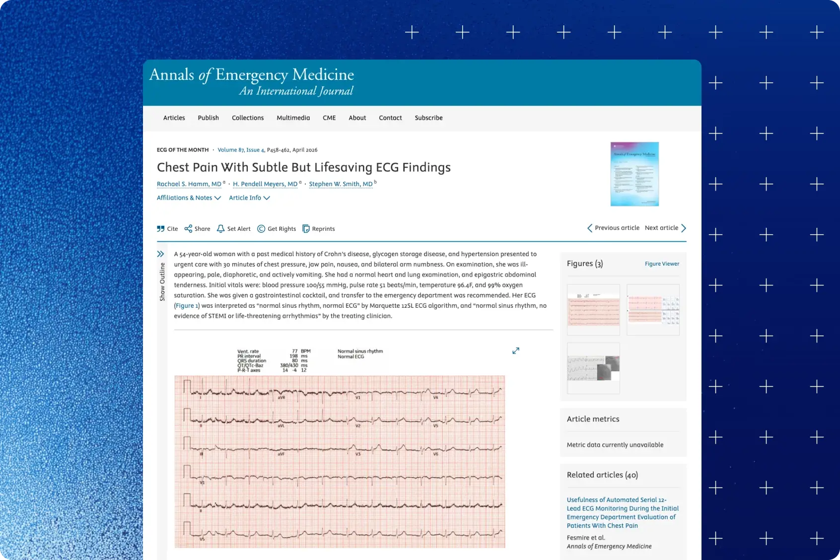
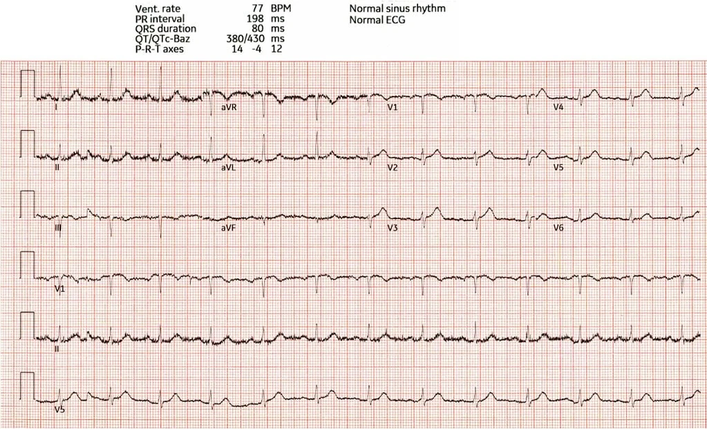
This study presents a clinical case highlighting the diagnostic value of hyperacute T waves on electrocardiogram (ECG) for early detection of acute coronary occlusion. A 54-year-old woman presented with classic ischemic symptoms, yet her initial ECG was interpreted as normal by both automated analysis and clinician review. Closer inspection revealed subtle hyperacute T waves in the anterior leads, consistent with acute occlusion of the left anterior descending (LAD) artery. The case underscores how reliance on traditional ST-segment elevation criteria alone may delay recognition of life-threatening myocardial infarction.
Key findings
- Case: 54-year-old woman, ischemic symptoms, initial ECG read as normal.
- Subtle anterior hyperacute T-waves indicated acute LAD occlusion.
- Reliance on ST-elevation criteria alone can delay treatment.
Published in: Annals of Emergency Medicine
Published on: 23 March 2026
Background
Timely identification of acute coronary occlusion is critical for initiating reperfusion therapy and improving outcomes. The current paradigm distinguishes ST-segment elevation myocardial infarction (STEMI) from non-STEMI (NSTEMI) based on ECG findings, particularly ST-segment elevation. However, this approach has important limitations: up to 25–30% of NSTEMI patients may have an unrecognized total coronary occlusion, associated with significantly higher mortality.
Hyperacute T waves have emerged as an early and specific ECG marker of occlusion myocardial infarction, often preceding ST elevation. Despite this, they are frequently overlooked due to their subtle appearance and lack of widespread familiarity or objective criteria. Recent work has introduced quantitative methods based on T-wave magnitude and symmetry, but these are not routinely applied in clinical practice.
Methods
This report describes a single-patient case analysis within the “ECG of the Month” educational framework. Clinical presentation, ECG findings, and subsequent clinical course were reviewed. The initial ECG obtained at urgent care was compared with the prior baseline ECG and follow-up ECGs during deterioration.
Key ECG features were analyzed qualitatively and in reference to established criteria for hyperacute T waves, including:
- Increased T-wave amplitude relative to the QRS complex
- Symmetric T-wave morphology
- Changes compared to baseline ECG
Clinical outcomes, including cardiac arrest, angiographic findings, and response to intervention, were documented to correlate ECG findings with definitive diagnosis.
Results
The patient presented with chest pain, nausea, and autonomic symptoms, but her initial ECG was interpreted as normal. Retrospective analysis demonstrated hyperacute T waves in leads V2–V5, with subtle (<1 mm) ST elevation.
Shortly after leaving urgent care, the patient suffered ventricular fibrillation cardiac arrest. Following resuscitation, the repeat ECG showed clear anterior STEMI. Coronary angiography confirmed acute thrombotic occlusion of the mid-LAD, which was treated with stenting. Despite intervention, the patient developed cardiogenic shock requiring advanced mechanical support, with severely reduced left ventricular ejection fraction (10%).
Comparison with baseline ECG revealed increased T-wave symmetry and magnitude, along with reduced QRS amplitude—features consistent with hyperacute T waves (illustrated on page 2).
Conclusion
This case demonstrates that hyperacute T waves can serve as an early and highly specific indicator of acute coronary occlusion, even in the absence of diagnostic ST-segment elevation. Failure to recognize these subtle ECG findings may delay life-saving treatment and worsen clinical outcomes. Greater awareness and education regarding hyperacute T waves—along with potential integration of automated or AI-based detection tools—may improve early diagnosis, guide urgent reperfusion decisions, and ultimately reduce morbidity and mortality in patients with acute coronary syndromes.
Authors: Rachael S. Hamm, MDa; H. Pendell Meyers, MDa; Stephen W. Smith, MDb
Share this article



About PMcardio
PMcardio is the market leader in AI-powered diagnostics, addressing the world’s leading cause of death – cardiovascular diseases. The innovative clinical assistant empowers healthcare professionals to detect up to 40 cardiovascular diseases. In the form of a smartphone application, the certified Class IIb medical device interprets any 12-lead ECG image in under 5 seconds to provide accurate diagnoses and individualized treatment recommendations tailored to each patient.

About Powerful Medical
Powerful Medical leads one of the most important shifts in modern medicine by augmenting human-made clinical decisions with artificial intelligence. Our primary focus is on cardiovascular diseases, the world’s leading cause of death.
Established in 2017, Powerful Medical has embarked on a mission to revolutionize the diagnosis and treatment of cardiovascular diseases. We are a medical company backed by 28 world-class cardiologists and led by our expert Scientific Board with decades of experience in daily patient care, clinical research, and medical devices. The results of our research are implemented, developed, certified, and brought to market by our 50+ strong interdisciplinary team of physicians, data scientists, AI experts, software engineers, regulatory specialists, and commercial teams.