
Supported Diagnoses
Validated across millions of ECGs. Used by 100,000+ clinicians worldwide.
PMcardio supports 50+ ECG findings — including conditions that are routinely missed without AI.
Available in:


Queen of Hearts™ (Myocardial Infarction)
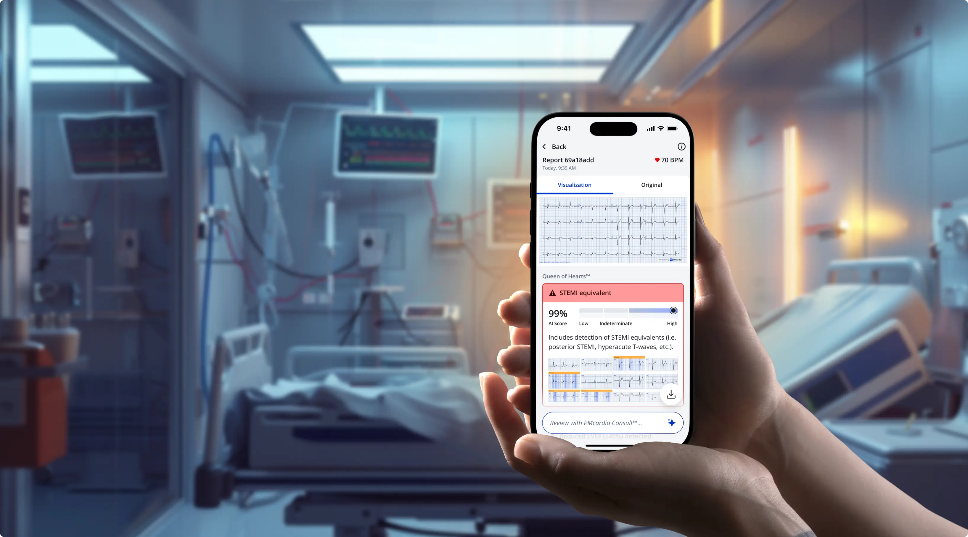
STEMI
STEMI Equivalent
Detects occlusive myocardial infarction (OMI) even without ST elevation (i.e. posterior STEMI, hyperacute T-waves, etc.). Negative for STEMI mimics (i.e. early repolarization, LVH, etc.)High-Risk NSTEMI (Reperfusion)
Represents a type 1 myocardial infarction caused by a transiently recanalized coronary occlusion—classically seen in patterns such as Wellens type A or B due to subtotal LAD obstruction, but possible in any infarct-related territory.High-Risk NSTEMI (Subendocardial Ischemia)
Detects subendocardial ischemia consistent with multivessel disease, partial coronary obstruction, or supply–demand mismatch. Explainability heat map is not yet available for this pattern.Culprit Detection
AI-predicted likelihood scores for LAD, LCx, and RCA with 3D heart visualization highlighting the predicted culprit artery.
LVsense™ (Left Ventricular Systolic Function)
Mildly reduced LVEF (41 – 49%)
No signs of reduced LVEF (≥50%)
Rhythms
Sinus bradycardia
Sinus rhythm
Sinus tachycardia
Paced rhythm
Atrial fibrillation
Atrial fibrillation with rapid ventricular response
Atrial fibrillation with slow ventricular response
Atrial flutter
Atrial flutter with rapid ventricular response
Atrial flutter with slow ventricular response
Supraventricular tachycardia
Junctional rhythm
Junctional bradycardia
Accelerated junctional rhythm
Wide QRS rhythm
Idioventricular rhythm
Wide QRS tachycardia
Conduction Abnormalities
1st degree AV block
2nd degree AV block, type Wenckebach
Higher degree AV block
Complete right bundle branch block
Incomplete right bundle branch block
Complete left bundle branch block
Incomplete left bundle branch block
Nonspecific intraventricular conduction delay
Left anterior fascicular block
Left posterior fascicular block
Bifascicular block (RBBB + LAFB)
Bifascicular block (RBBB + LPFB)
Trifascicular block (RBBB + LAFB + AVBLOCK1)
Trifascicular block (RBBB + LPFB + AVBLOCK1)
Other Supported Conditions
Atrial enlargement
Long QT syndrome
Short QT syndrome
Ventricular hypertrophy
Premature complexes
Axis
Left cardiac axis deviation
Right cardiac axis deviation
Extreme cardiac axis deviation
Normal axis
Measurements
Heart rate
P wave
PR interval
QRS duration
QT interval
Corrected QT interval (Framingham)
Corrected QT interval (Bazett)
RR interval
PP interval
ST elevations
Hyperacute T-waves (HATW)
Research Models Available
for Selected Partners and Internal Use.
Available in:
Serum Potassium Prediction (Hyperkalemia and Hypokalemia)
Median Beats
Serial ECG Comparison
Advanced ECG Measurements (>100 fiducial points)
ECG Signal Quality Indicators
Lead Reversal Detection
Acute Right Heart Strain (including pulmonary embolism-associated presentations)
Electrolyte imbalances (including Hypercalcemia, Hypocalcemia, Hypermagnesemia, and Hypomagnesemia)
Currently Unsupported Diagnoses
Asystole
Sino-Atrial Exit Block (and its different types)
Sick sinus syndrome
Sinus pause
Digitalis effect or intoxication
Pericarditis
Pericardial effusion/tamponade
Myocarditis
Brugada Syndrome
Hypothermia (Osborn wave)
Arrhythmogenic cardiomyopathy (ACM, arrhythmogenic right ventricular cardiomyopathy (ARVC) – epsilon waves)
Dextrocardia
Pre-excitation syndromes (Wolff-Parkinson-White [WPW] Syndrome
Any other not explicitly supported diagnosis
Proven Impact.
Measurable Results.
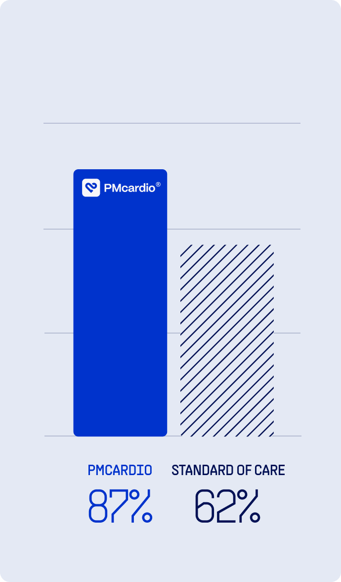
See how PMcardio outperforms standard care in real clinical settings with up to 2× higher sensitivity in detecting heart attacks. Validated in 25+ external, independent studies.


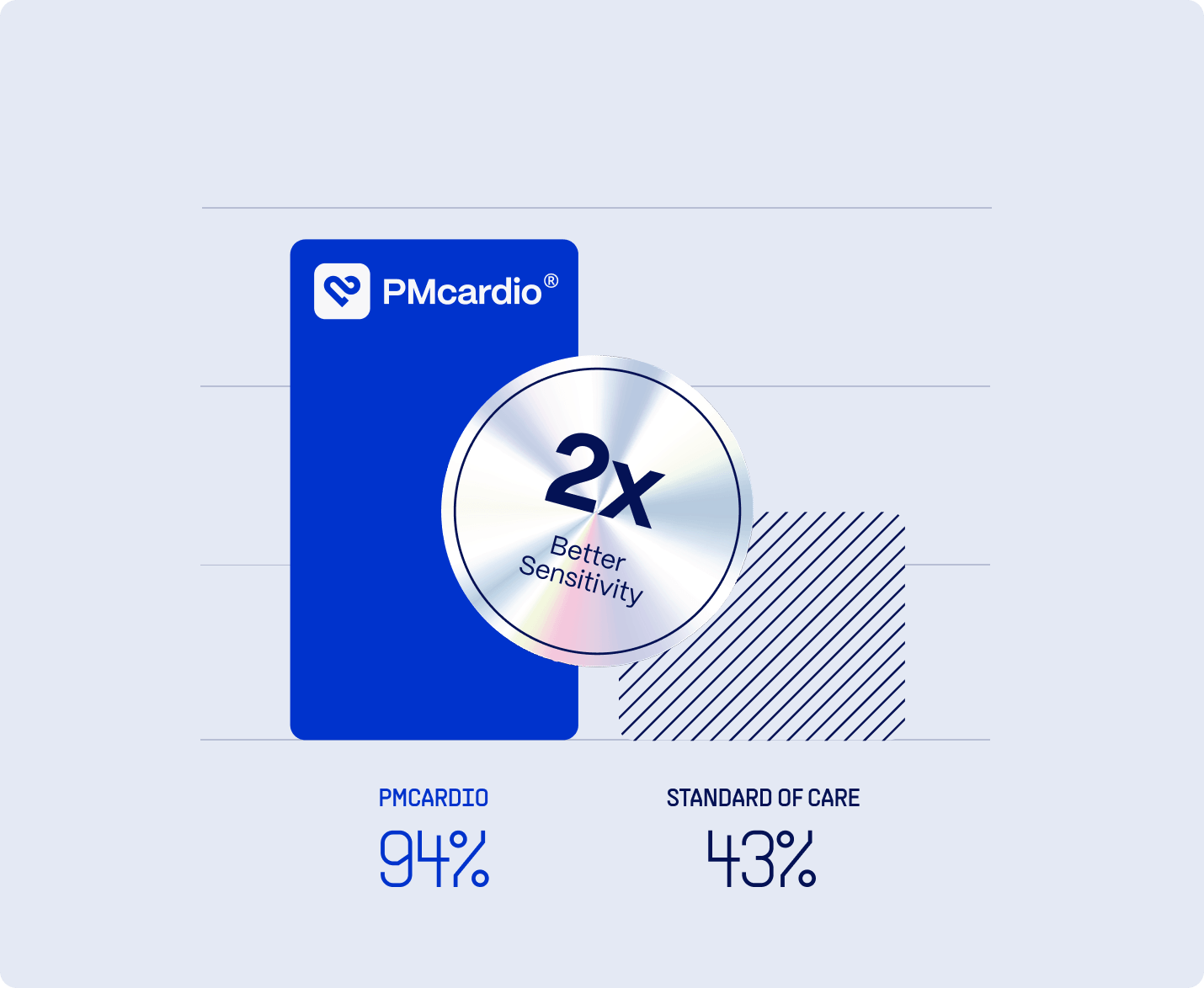
Heart Attacks
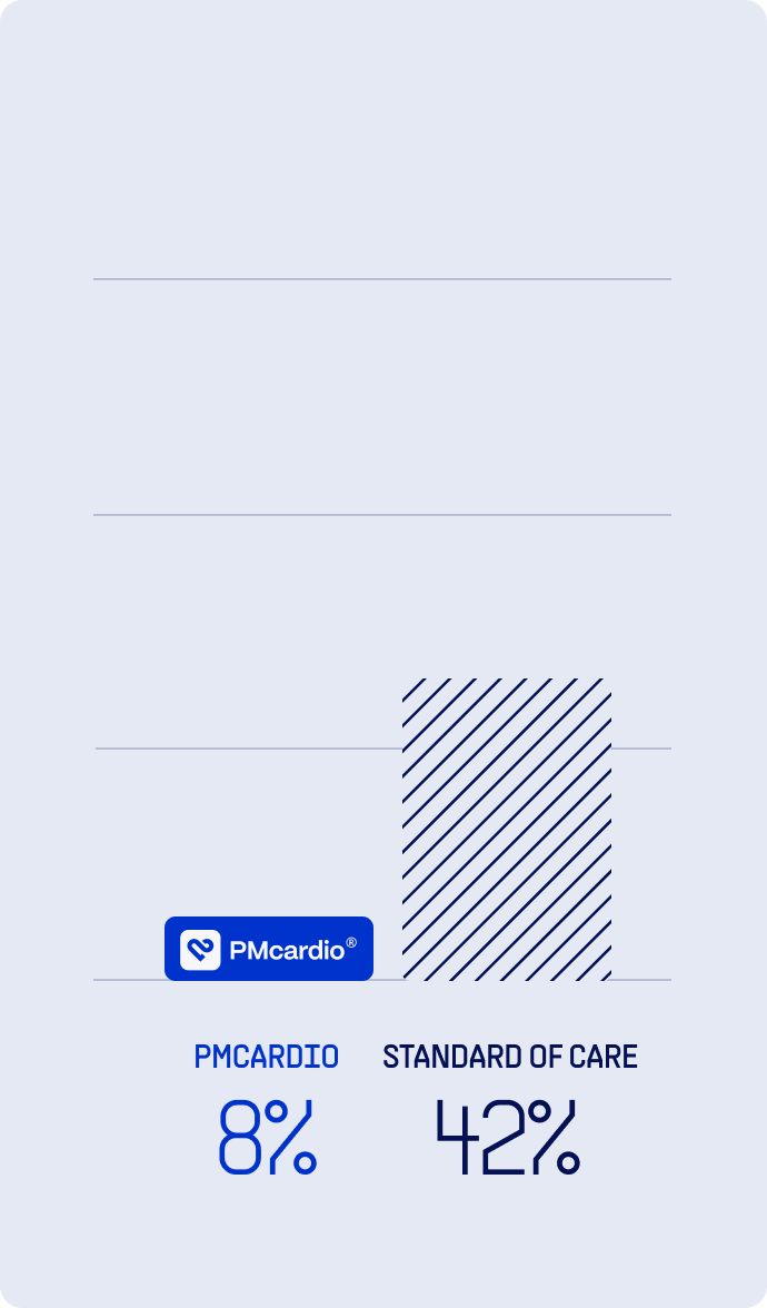
False Positives
Heart Blocks
PMcardio Provides Direct Clinical and Economic Benefits within just 6 months of implementation
Try PMcardio
for Free
Join 100,000+ clinicians already using PMcardio to detect heart conditions with AI-powered accuracy. Get instant access to PMcardio, designed for speed, simplicity, and clinical confidence.


- No credit card required
- 5 FREE ECGs/month
- Works with any 12-lead ECG image