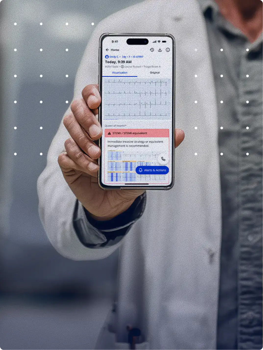
Get instant AI-powered ECG interpretations anytime, anywhere. Trusted by over 100,000 healthcare professionals
PMcardio puts instant AI-powered ECG analysis into clinicians' hands — from emergency departments and ambulances to clinics and hospital wards — helping them make faster, more confident decisions at every point of care.
How it works
3 Simple Steps to Instant ECG Interpretation
Step 01.
Snap a Photo of Any 12-lead ECG
Capture an ECG from paper, screen, or upload an image from your gallery.
Step 02.
Detect 50+ ECG Findings in Seconds
Accurately detect STEMI/OMI, rhythms, reduced LVEF, conduction disorders and other ECG abnormalities.
Step 03.
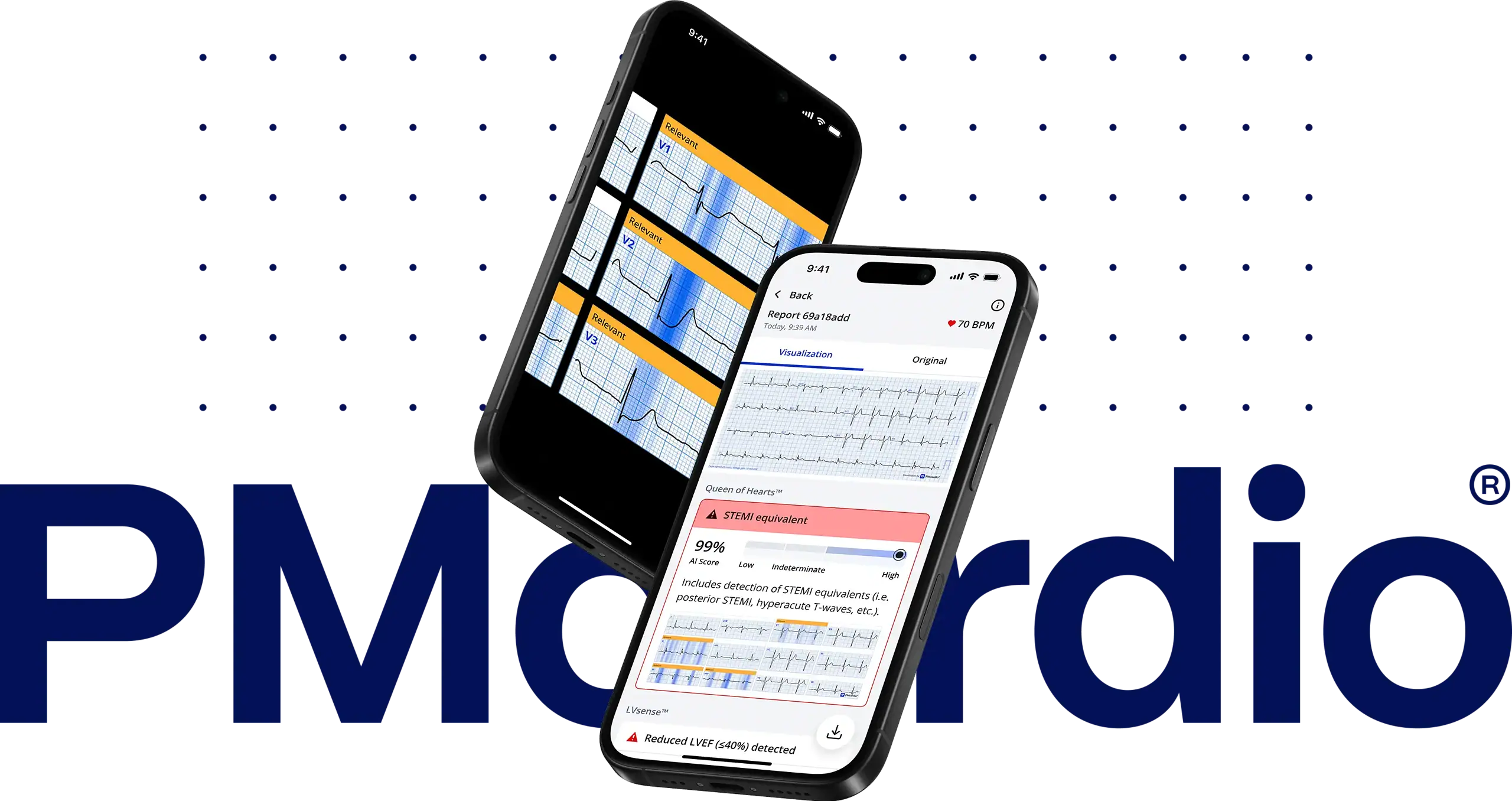
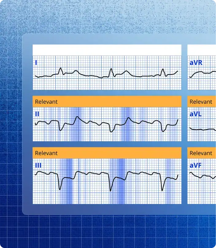
Visualize Results with Explainability Heatmaps
See which features matter most so you can interpret ECGs with confidence.
SPOTLIGHT
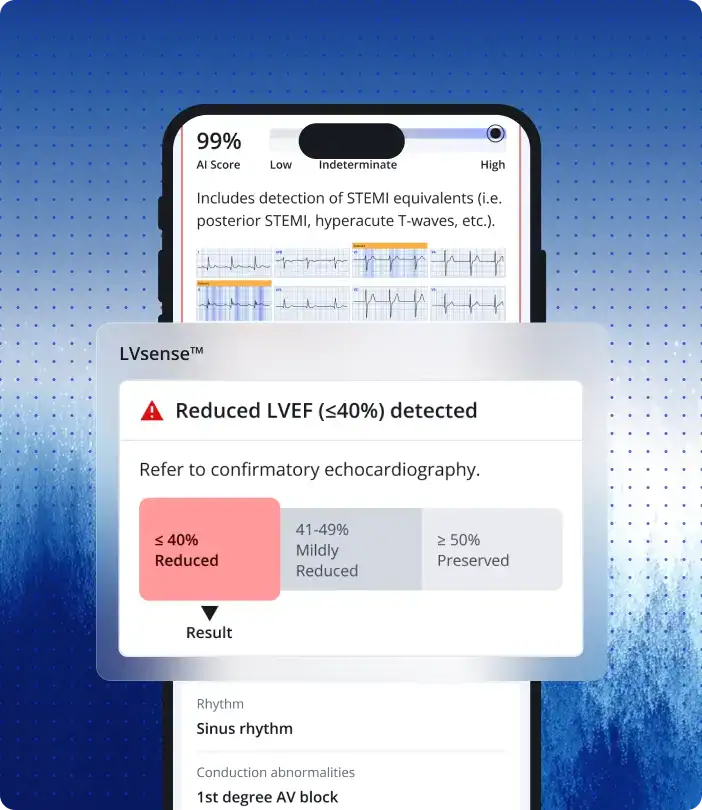
Early Detection of Reduced LVEF on ECG with LVsense™
Rapid and accurate assessment of left ventricular (LV) systolic function.
SPOTLIGHT
Identify Every Clinically Relevant ECG Finding
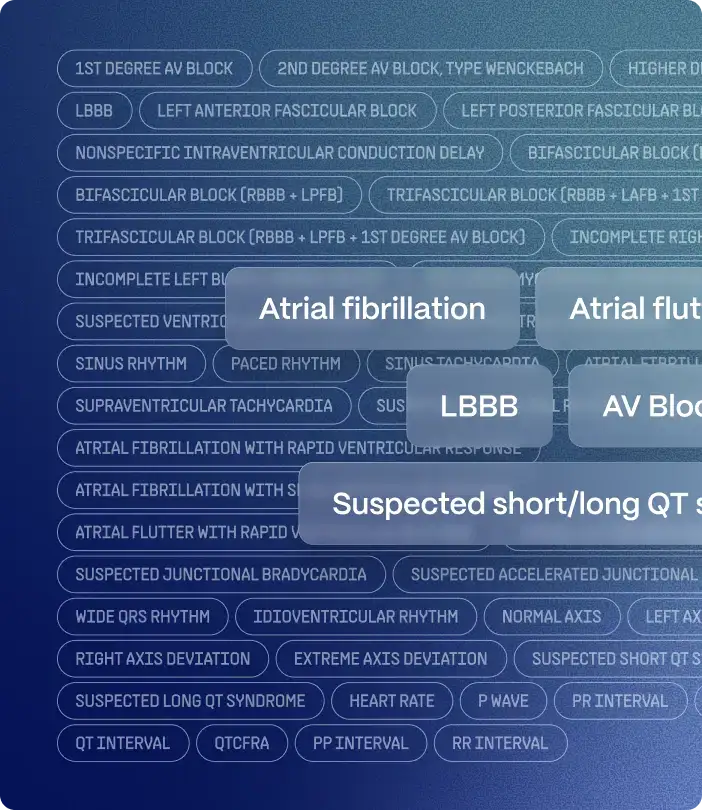
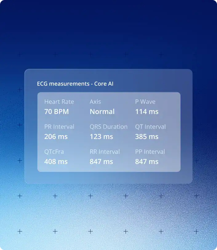
From atrial fibrillation and conduction disorders to LV function screening and precise interval measurements — every finding is structured and ready to act on. No interpretation left to chance.
120K
Heart Attacks Detected
100,000+
Clinicians Worldwide
2.5M+
ECGs Analyzed
25+
Clinical Validation Studies
150+
Hospital Deployments
Start reading ECGs with AI — free on iOS and Android.
Benefits
Why 100,000+ Clinicians Trust PMcardio
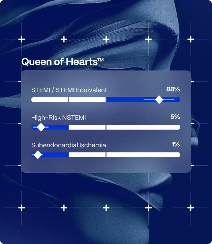
Queen of Hearts™ - STEMI and OMI Detection
Detect occlusive myocardial infarctions (OMIs) even in the absence of ST elevation, while reducing false positives from common STEMI mimics.
Detect reduced left ventricular ejection fraction (LVEF) directly from the ECG, enabling early identification of systolic dysfunction before symptoms progress.
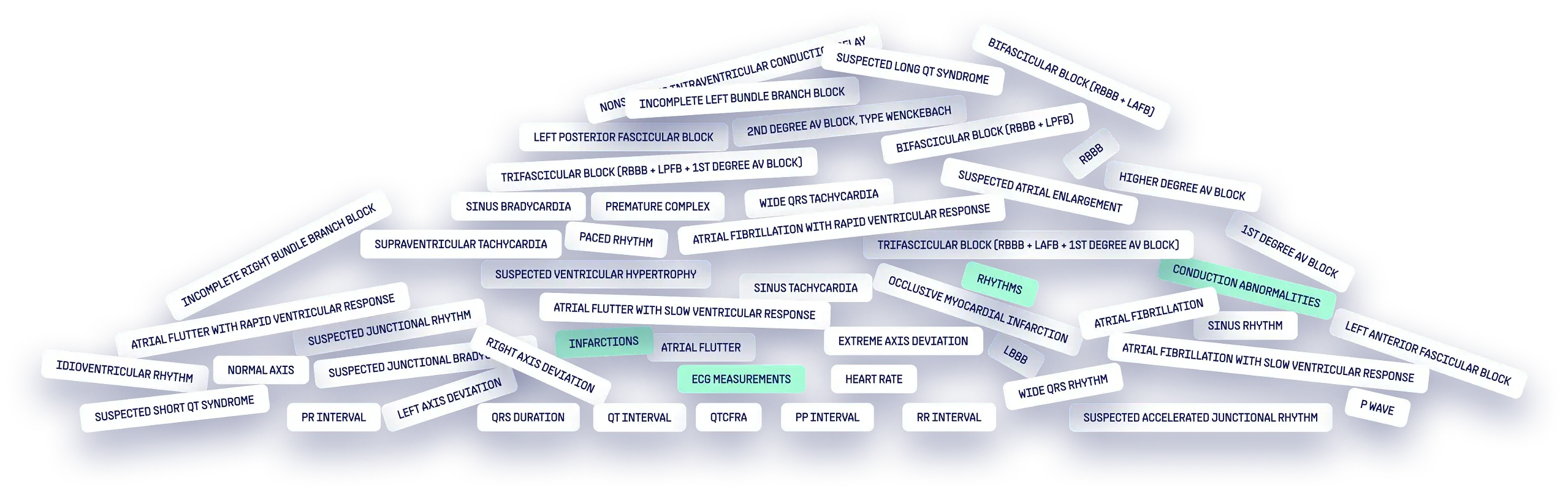
Covers 50+ ECG findings across rhythms, conduction disorders, structural changes, and acute ischemia — including conditions that are routinely missed without AI.
Trained on large, geographically and ethnically diverse datasets with validated angiographic outcomes — built to perform across all patient populations.
heart blocks
infarctions
ectopies
hypertrophies
rhythms
arrhythmias
axis
measurements
others
Clinical Evidence
Proven Impact.
Measurable Results.
See how PMcardio outperforms standard care in real clinical settings with up to 2× higher sensitivity in detecting heart attacks. Validated in 25+ external, independent studies.
PMcardio Provides Direct Clinical and Economic Benefits within just 6 months of implementation
“PMcardio’s STEMI AI ECG Model leverages decades of our ECG morphology research to accurately distinguish acute occlusions from patterns which mimic them, going beyond mere ST-elevation analysis.”
Prof. Stephen W. Smith, MD Emergency Physician Hennepin County Medical Center Founder of Dr. Smith’s ECG Blog
“After integrating PMcardio, we noticed a clear drop in false cath lab activations. The AI accurately flags occlusions while minimizing noise, which has saved time, resources, and spared patients unnecessary procedures.”
“I feel so passionately that we should embrace the opportunity and adopt innovation like PMcardio that gives near-perfect accuracy for detecting diagnoses such as Atrial Fibrillation in primary care.”
Dr. Yassir Javaid Cardiovascular Clinical Lead East Midlands Clinical Networks, Clinical Director for Cardiology at Bupa
FAQ
Frequently Asked Questions
What is PMcardio?
PMcardio offers CE-certified AI ECG Models that read ECGs and offers a complex assessment of 50+ ECG findings. Clinically validated in 25+ studies and trusted by over 100,000 clinicians, it delivers rapid, expert‑level interpretations, empowering emergency physicians, GPs, nurses, paramedics, and cardiologists to act with confidence at the point of care.
Certain AI ECG Modules are CE-marked medical devices under EU MDR and only certified for marketing in the European Union and the United Kingdom. Powerful Medical technology has not yet been cleared by the US Food and Drug Administration (FDA) for marketing in the USA. Not all modules of the PMcardio platform may be available in your region.
Is it safe to upload ECG images to PMcardio?
Yes, your encrypted data never leaves your region. PMcardio is fully GDPR and HIPAA compliant and adheres to the strictest regulatory and security frameworks, including SOC 2 and ISO 27001 certifications.
For a detailed overview of our compliance, data protection practices, and security protocols, visit our Trust Center.
Is PMcardio clinically validated?
Yes, PMcardio’s AI ECG Models have been rigorously validated in more than 25 independent, peer‑reviewed clinical studies. Across these studies, PMcardio AI ECG interpretation outperformed standard‑of‑care methods, delivering:
2x sensitivity for early STEMI detection (often on the very first ECG)
Reduces false STEMI alarms by over 60%, preventing false positive cath lab activations
Identifies atrial fibrillation (and other arrhythmias) with 98.3 % accuracy – on par with board‑certified cardiologists
For more information, the latest published evidence, and ongoing RCTs, please visit our Research page.
Which ECG formats does PMcardio accept?
PMcardio supports every commercially available 12-lead ECG, whether as a single-page image or a multi-page report.
The only hard requirement is that the ECG (including all its leads) must be printed on electrocardiogram graph paper, where every five small (1 mm) squares, you can find a heavier line forming a larger 5 mm square — this is essentially the standard ECG paper. The entire ECG should appear on one continuous grid, meaning it should not be split across multiple intersecting grids. Additionally, the image must contain only the relevant leads, without any leads from other ECGs intruding into the picture.
Fewer-lead ECGs, such as 1-lead, 3-lead, or 6-lead recordings, are currently not supported.
What ECG abnormalities can PMcardio detect?
PMcardio’s AI-powered ECG algorithms recognise 50+ ECG findings across four major categories:
Is PMcardio just another AI chatbot like ChatGPT or Perplexity?
No. PMcardio’s AI ECG Models are deterministic, CE-certified medical devices, purpose-built for ECG interpretation, not a conversational large language model (LLM).
Zero hallucinations: The same ECG file always returns the same diagnosis.
Evidence-based training: Fixed model weights are derived from 4 million+ clinically labelled ECGs with matched patient outcomes.
Independent proof: Performance has been independently validated in 25+ peer-reviewed validation studies across Europe and the U.S.
Regulatory oversight: Post-market surveillance and quality-management systems ensure ongoing compliance with Medical Device Regulation.
Learn more about how PMcardio compares to AI chatbots like ChatGPT.
Is PMcardio more accurate than automated ECG machine reads?
Yes. In six peer-reviewed studies, PMcardio’s AI ECG algorithms has demonstrated up to 2x higher accuracy than conventional ECG algorithms used in standard ECG machines (e.g., GE Marquette 12SL, Philips, Glasgow Algorithm).
Learn more about how PMcardio compares to Conventional ECG Algorithms.
Is PMcardio free?
Yes. Start with a Free Plan that lets you analyse up to 5 ECGs per month at no cost. If you need more reports and advanced Pro features, you can upgrade to a paid subscription with:
Larger monthly ECG quota: run as many studies as your workflow demands.
AI explainability for STEMI: colour-coded highlights show the waveform segments driving each positive diagnosis.
12 validated interval measurements: instant PR, QRS, QTc intervals and more.
30-day report history: revisit, download, or share any ECG analysed in the past month.
* Certain AI ECG Modules are CE-marked medical devices under EU MDR and only certified for marketing in the European Union and the United Kingdom. Powerful Medical technology has not yet been cleared by the US Food and Drug Administration (FDA) for marketing in the USA. Not all modules of the PMcardio platform may be available in your region.
PMcardio® For Organizations
Your Hospital Runs on ECGs.
Make Every One Count.
PMcardio for Organizations brings AI ECG interpretation into your institution’s clinical workflow — with team management, device integrations, and real-time alerting built for hospitals, EMS, and health systems at scale.
Institution-Wide ECG Access
Every ECG across every location, visible in one admin dashboard. No per-seat limits, no data silos.
Direct ECG Device & EHR Integration
Connect existing ECG machines and hospital systems. Results flow automatically, no manual uploads.
Real-Time Alerts & Care Routing
Instant notifications when a critical finding is detected, with built-in escalation to the right team member.
Schedule a Demo
Talk to our team and get a walkthrough tailored to your department, devices, and clinical setup.
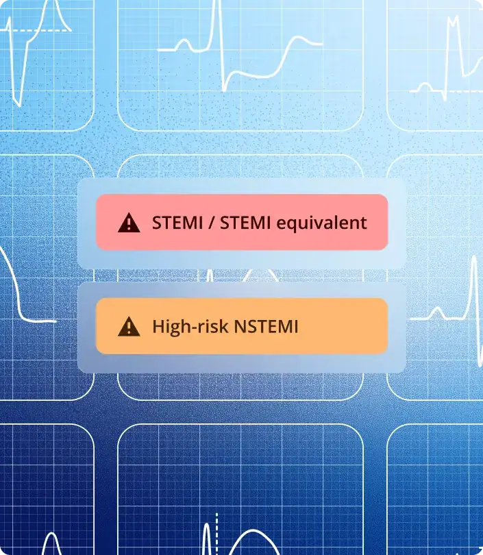
STEMI Equivalent Equivalent Detects occlusive myocardial infarctions (OMIs) even without ST elevation (i.e. posterior STEMI, hyperacute T-waves, etc.). Negative for STEMI mimics (i.e. early repolarization, LVH, etc.)
High-Risk NSTEMI Represents a type 1 myocardial infarction caused by a transiently recanalized coronary occlusion—classically seen in patterns such as Wellens type A or B due to subtotal LAD obstruction, but possible in any infarct-related territory.
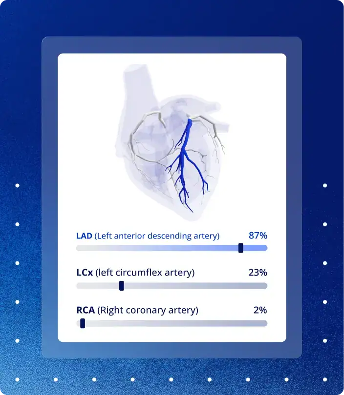
Culprit Detection AI-predicted likelihood scores for LAD, LCx, and RCA with 3D heart visualization highlighting the predicted culprit artery.
Conduction Abnormalities (Heart Blocks 1st degree AV block • 2nd degree AV block, type Wenckebach • Higher degree AV block • Complete right bundle branch block • Incomplete right bundle branch block • Complete left bundle branch block • Incomplete left bundle branch block • Nonspecific intraventricular conduction delay • Left anterior fascicular block • Left posterior fascicular block • Bifascicular block (RBBB + LAFB) • Bifascicular block (RBBB + LPFB) • Trifascicular block (RBBB + LAFB + AVBLOCK1) • Trifascicular block (RBBB + LPFB + AVBLOCK1)
LVEF Reduced LVEF (≤40%) • Mildly reduced LVEF (41 – 49%) • No signs of reduced LVEF (≥50%)
Axis Left cardiac axis deviation • Right cardiac axis deviation • Extreme cardiac axis deviation • Normal axis
Other Supported Diagnoses Suspected long QT syndrome • Suspected short QT syndrome • Suspected atrial enlargement • Suspected ventricular hypertrophy • Premature complexes
Certain AI ECG Modules are CE-marked medical devices under EU MDR and only certified for marketing in the European Union and the United Kingdom. Powerful Medical technology has not yet been cleared or approved by the US Food and Drug Administration (FDA) for marketing in the USA. Not all modules of the PMcardio platform may be available in your region.