Powerful Medical Receives €40 Million IPCEI Grant — read the full story

Prof. Dr. Robert Hatala, PhD
2. March 2023

Athletes Collapsing: A Deep Dive into the Heart’s Electrical Chaos and the Fight for Survival

The sudden collapse of a top athlete in the middle of a game, often on live TV cameras, is usually accompanied by questions about the cause of this dramatic situation. The public is mostly mistakenly convinced that the athlete had a heart attack. However, the truth is that heart attacks rarely cause these life-threatening situations in young people.

The cause of the dramatic collapse of top athletes (but of course not only them), usually within seconds, is a severe sudden disturbance in the heart’s electrical activity – arrhythmia.

Why are young athletes collapsing: Understanding Arrhythmia and Heart’s Electrical Disturbances

Arrhythmia is the creation of very fast chaotic electrical impulses reaching a frequency of around 300-400/minute, during which the pumping action of the heart stops, the blood stops flowing into the body, and we, therefore, speak of circulatory arrest. The brain is the first organ to react to this potentially fatal situation, and the patient loses consciousness.

Case Study: Damar Hamlin’s Near-Fatal Collapse

Even earlier this year, 24-year-old Buffalo Bills safety Damar Hamlin suddenly collapsed in the middle of an American football game. Following a routine tackle, he went into cardiac arrest. Thanks to the timely deployment of a defibrillator, he survived the arrhythmia and is on the mend now.

But why did it happen? There is no straightforward explanation – the occlusion of circulation can have many causes. Let us look at what we know about the heart’s electrical activity and how situations like the one happening to Mr Hamlin could be prevented.

The chaos of the heart’s electrical impulses can be deadly.

The heart is a muscular pump that pumps blood in parallel through 2 circuits – the “large” whole-body circuit and the “small” pulmonary circuit. The heart muscle alternately contracts and relaxes, sucking deoxygenated blood from the body and pushing it through the lungs through the right ventricle. After oxygenation, the blood returns to the left ventricle, which ejects it into the aorta and, from there, to the whole body.

A correctly timed electrical impulse conditions the correct function of the heart and its every contraction. Physiological electrical impulses arise in a cluster of special cells in the confluence of a large vein from the upper part of the body (the so-called superior vena cava) and the right atrium of the heart, which is called the sinus node.

The Electrical Symphony of the Heart

The sinus node creates electrical impulses with a frequency corresponding to the need for sufficient blood supply for the whole organism. This need is very variable and, depending on the load and the level of neurovegetative stimulation, it usually ranges from 50 to 200 impulses per minute in a young, healthy organism.

The electrical impulse is then distributed through a network of special cells that function as cables to all areas of the heart muscle. This activity ensures the synchronous contraction of the ventricles, which is necessary for optimal efficiency in expelling blood from the heart.

Athletes Collapsing: A Deep Dive into the Heart's Electrical Chaos and the Fight for Survival

Stay on the pulse with our newsletter

Your submission was successful

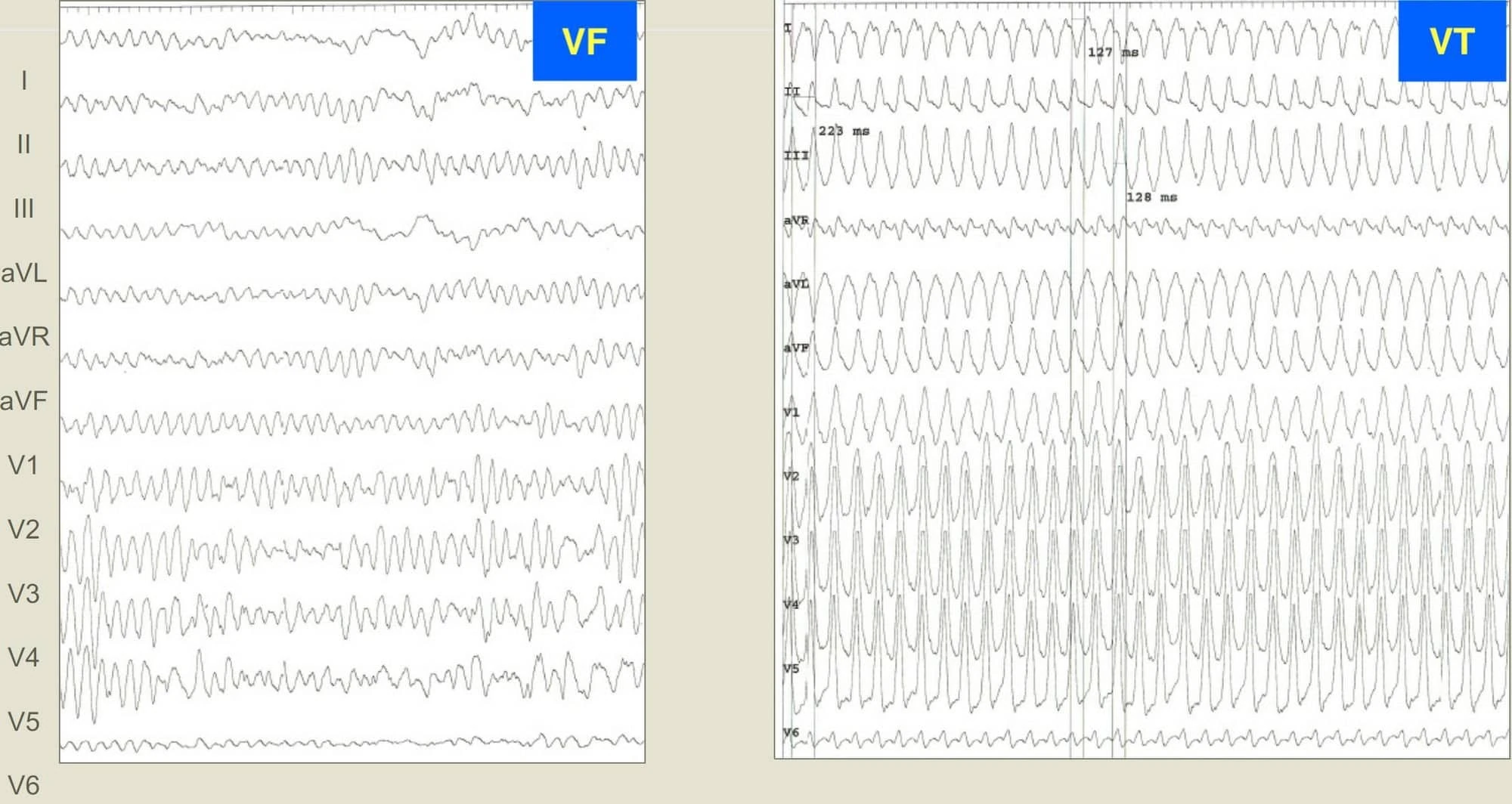
Ventricular Fibrillation: A Lethal Arrhythmia

One of the disturbances in the heart’s electrical activity is the sudden appearance of several deposits in the muscle of the heart chambers, which begin to send uncoordinated high-frequency electrical impulses to their surroundings – 300-400 times per minute.

As a result, the heart begins to oscillate chaotically and immediately stops pumping blood – blood circulation stops. This type of malignant arrhythmia is called ventricular fibrillation – the most common cause of sudden cardiac death.

Note that ventricular fibrillation fundamentally differs from atrial fibrillation, a common, often asymptomatic arrhythmia affecting 3% of the adult population.

If the ventricular fibrillation is not treated within a few minutes, irreversible damage occurs, and gradually all organs fail. First is the brain, the most sensitive organ to oxygen deprivation.

Athletes Collapsing: How Can a Fatal Pathological Process Be Reversed?

Averting the fatal end of someone suffering from ventricular fibrillation presupposes restoring the correct electrical function of the heart as soon as possible – i.e., eliminating electrical chaos. Applying an electrical discharge with several hundred volts and a short millisecond duration can achieve this most effectively.

Such a discharge induces an electrical “reset” of the heart – the entire heart contracts at once with a powerful discharge. There is a brief electrical silence, and regular electrical activity is restored with the return of blood circulation.

Reversing the Fatal Process: The Role of Defibrillation

However, to apply such an “electric shock,” it is necessary to have a special device – a defibrillator.

Ideally, a defibrillator should be brought to a victim of ventricular fibrillation within a few minutes. Since a defibrillator is typically available at the sports field, the chance to save an athlete who suffered cardiac arrest during a sporting event is generally relatively high. In real life, waiting for a defibrillator usually takes much longer – often tens of minutes after which the chance for patient survival is minimal.

While waiting for a defibrillator, a patient must be given proper resuscitation. Their survival depends on it. After securing the airways (preventing the tongue-swallowing), its essence is a powerful rhythmic chest compression at the sternum level with both palms placed on top of each other.

The Crucial Window for Action

The compression speed should be about 100 compressions per minute – this is approximately the rhythm of the well-known Bee Gees song with a very fitting name, “Stayin’ Alive”. With correctly applied compression of sufficient force, a rib or ribs may break (often audibly), but it is a negligible complication in this situation.

If an electric defibrillation shock with the restoration of blood circulation is timely applied, bridged by proper resuscitation, we are talking about an averted sudden death. In general, without providing resuscitation manoeuvres, the victim’s probability of survival decreases by 10% with each passing minute, so after 10 minutes of untreated ventricular fibrillation, the patient usually dies.

Several scientific works have convincingly proven that correct and timely resuscitation by laypeople by witnesses of the event is a decisive factor in averting sudden death and surviving the patient without brain damage.

Athletes Collapsing: A Deep Dive into the Heart's Electrical Chaos and the Fight for Survival

Try PMcardio for Free​

Join 100,000+ healthcare professionals using the world’s most advanced ECG reader app. Experience faster, smarter cardiac care—starting with 5 FREE ECGs/month. No credit card required.

A fight against time

After a nationwide campaign on proper resuscitation in Denmark, the survival rate of patients after sudden circulatory arrest increased 4-fold to more than 30%.

However, first aid must be completed as quickly as possible using an automated external defibrillator (AED). These are most often located in places with a high density and movement of people (transport hubs, sports fields, shopping centres, hotel lobbies, etc.).

Delayed arrival of the emergency medical service, whether due to late contacting its dispatch or due to the poor condition of the road network and heavy traffic in cities, can be fatal for the patient.

In some areas of Sweden, trials have begun to use drones controlled by the rescue centre to transport the defibrillator to the victim. However, for the rescue operation to start at all, it is vitally important to immediately inform the dispatch of the rescue service about the case.

Despite timely delivery of defibrillation, circulation cannot always be successfully restored, and the heart fails to return to normal electrical function. In these situations, highly specialised assistance with oxygen administration, airway intubation, intravenous drug administration, or extracorporeal circulation support and cooling of the patient is required.

Why ventricular fibrillation occurs – young heart vs. old heart

The causes of sudden arrhythmic death vary greatly, depending on the patient’s age. However, sudden death at a young age is, for understandable reasons, perceived more dramatically than in a person who has been receiving heart treatment for years.

For a layperson, it may seem incomprehensible how such a severe condition can occur in a person representing the very symbol of physical health – a top athlete – who also undergoes periodic preventive medical examinations by sports doctors.

However, such life-threatening sudden events with much media attention are relatively rare, occurring in approximately one in 15,000 athletes per year, significantly less than in the “non-athletic” population.

From a global perspective, sudden cardiac death accounts for approximately 15% of all deaths, a significant mortality rate.

The conducted studies of victims of sudden cardiac death show two types of diseases that young people, including athletes, typically suffer from:

The first group of diseases

Primary electrical diseases of the heart are most often a result of genetic mutations, of which we still know only a fragment. Gene mutations cause, under certain circumstances, abnormal permeability of ions through the membranes of some heart cells, which causes the abnormal spontaneous electrical activity of these cells, which sets off “electrical fireworks” in the heart.

The malfunction can only occur under certain circumstances, e.g. during extreme heart stimulation with adrenaline, under the influence of doping, at a high environmental temperature (e.g. a hot bath), or after using drugs.

Such a genetic disorder cannot be detected during a routine preventive examination. An electrocardiogram (ECG) at rest and during exercise and an ultrasound examination of the heart can be completely normal. However, essential data towards the correct diagnosis can be, e.g., sudden death under the age of 50 in blood relatives or dropping out in childhood.

Athletes Collapsing: A Deep Dive into the Heart's Electrical Chaos and the Fight for Survival

Read your next ECG with certified AI

The second group of diseases

The primary genetically determined disease of the heart muscle is the so-called hypertrophic cardiomyopathy. At a young age, this can be manifested only by a relatively insignificant thickening of the heart’s walls, which, however, also occurs physiologically in athletes as a result of their high level of training. A genetic examination supplemented by a magnetic resonance heart examination can be decisive.

Younger age groups: The case of an American football player

When it comes to Damar Hamlin, the above-mentioned football player, what was probably the case was an underestimated mechanism of arrhythmia – the player collapsed after a sudden collision with another player who hit him in the chest with his helmet while running.

When a hard object strikes the chest in sports, such as a baseball or basketball, the mechanical energy of the impact can be converted into an electrical impulse, which can disrupt the heart’s rhythm and trigger a fatal arrhythmia, even in those with perfectly healthy hearts.

This occurrence is the so-called ‘concussion of the heart’, in Latin commotio cordis, a somewhat archaic name still used in some professional literature. This mechanism can also be triggered when the chest hits the steering wheel in a traffic accident.

Higher age groups

In the older-adult category (over 50 years of age), the most common cause is acute lack of blood supply to the heart muscle – ischemia – due to the closure of the blood flow in the large arteries that supply the heart. Atherothrombosis is typically the reason for artery closure. Fat deposits cause an artery’s narrowing; subsequently, a blood clot forms and blocks the vessel like a plug. In some cases, arrhythmia – ventricular fibrillation – occurs within seconds, which kills the patient even before the heart attack develops, i.e. the heart muscle dies.

Nowadays, most patients survive a heart attack but are left with a scar of varying sizes on their hearts. Such a chronic scar is the most common cause of a slightly different but potentially fatal arrhythmia in people after a heart attack – the arrhythmia is called ‘ventricular tachycardia‘.

In ventricular tachycardia, the impulses are initially more organised than in ventricular fibrillation and usually have a lower frequency, typically in the range of 160-240 impulses per minute. The patient does not always lose consciousness immediately. However, without treatment, such an arrhythmia usually degenerates into chaotic ventricular fibrillation with a life-threatening outcome within a few minutes. Timely use of the defibrillator is again crucial for the patient’s survival.

In the case of post-infarction arrhythmias, ventricular tachycardia triggers an electrical impulse that “circulates” in the scar tissue caused by the infarction. In this case, we can suppress the arrhythmia by successfully “burning” this part of the scar with a catheter through which we apply a high-frequency electric current (microwave principle). This method is referred to as catheter ablation.

The method can also be used in patients in whom an abnormal electrical impulse (which always originates from one or a few cell clusters on the inner surface of the heart’s chambers) causes ventricular fibrillation. By eliminating these cell clusters, we remove the arrhythmia trigger.

Two sample ECGs, one depicting ventricular fibrillation, the other depicting ventricular tachycardia
Image 1: An ECG of ventricular fibrillation (left) shows a chaotic, irregular electrical activity in which the heart seemingly helplessly flutters. As a result, it ceases to pump blood. An ECG of ventricular tachycardia (right) shows fast, regularly following flutters with a frequency of 280/min. These flutters arise in the muscle of the ventricles. Although less chaotic than in VF, they may also cause blood circulation to stop if they persist longer.

Untreated high blood pressure

A heart damaged by untreated high blood pressure is also a critical terrain for these arrhythmias. With increasing age, the heart – apart from ageing – is often simultaneously affected by several pathological processes, significantly increasing the risk of sudden cardiac death.

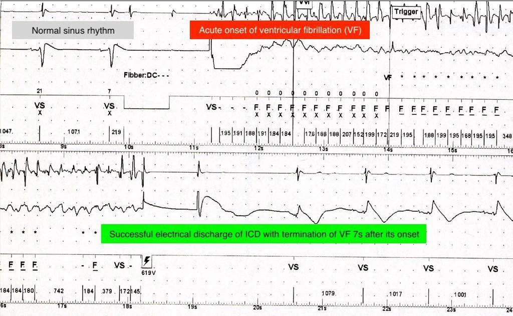
In current-state cardiology, it is possible to identify some patients with such a risk and implant a special generator of electric impulses – a defibrillator – under the skin in the chest area. It is a compact device in shape and size, similar to a classic pocket watch, weighing around 70 grams. Here’s schema of an implantable defibrillator (ICD) in the area below the left collarbone with electrodes inserted into the heart:

Athletes Collapsing: A Deep Dive into the Heart's Electrical Chaos and the Fight for Survival
Image source

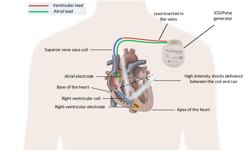
The device continuously monitors the electrical activity of the heart. In ventricular tachycardia or fibrillation, the device recognises the arrhythmia and terminates it within a few tens of seconds with a series of electrical impulses or a discharge of 40-80J (Image 2).

An ECG tracing showing how an implantable cardiac defibrillator (ICD) can successfully terminate a potentially fatal ventricular fibrillation
Image 2: A record from the implanted defibrillator (ICD – implantable cardioverter defibrillator) captures the moment of onset of ventricular fibrillation (VF), which the device almost immediately identifies as a life-threatening event. After 7 seconds of duration of the arrhythmia, the ICD successfully terminates it with an electrical discharge, restoring the heart’s normal electrical activity.

Can the potentially fatal collapses of athletes be prevented?

It is practically impossible to carry out preventive, highly specialised and expensive genetic and imaging examinations in millions of otherwise healthy people who want to do sports.

Although sudden cardiac arrests due to arrhythmia are rare in athletes compared to the rest of the population, they always attract the media’s attention and, thus, the entire public. Unfortunately, as it often happens, the first and only manifestation of the disease is a collapse in the sports field. If the sportsman/sportswoman survives the sudden cardiac death, the current special field of cardiology – arrhythmology – can help them further.

A real-life example is the case of 29-year-old Danish football player Christian Eriksen, who survived sudden cardiac arrest during an international match in 2021 and today, with an implanted defibrillator, continues his career as a football player for Manchester United.

Despite periodic examinations of top athletes, we are unlikely to detect all at-risk individuals in time in the foreseeable future. Sadly, this is why we will probably keep witnessing such dramatic cases.

However, the explosive development of artificial intelligence brings some optimism. First, in the advanced cardiac screening of large populations. Second, in analysing the electrocardiogram (ECG) using deep learning (deep learning is a form of machine learning using computer neural networks).

An AI-powered ECG analysis can identify previously undetected or overlooked content of an ECG recording, providing an unprecedented opportunity for early detection of the risk of developing life-threatening arrhythmias.

Co-founder and Chief Scientist

Prof. Dr. Robert Hatala, PhD

As the Head of the Arrhythmology Department at the National Institute of Cardiovascular Diseases, Prof. Dr. Hatala has contributed significantly to medicine with over 150 publications. He has been instrumental in shaping the European Society of Cardiology’s clinical guidelines and currently serves as Powerful Medical’s Chief Scientist.
About PMcardio:

PMcardio is a CE-certified AI that reads ECGs and offers a complex assessment of 49 cardiac conditions. Clinically validated in 15+ studies and trusted by over 100,000 clinicians, it delivers rapid, expert‑level interpretations, empowering emergency physicians, GPs, nurses, paramedics, and cardiologists to act with confidence at the point of care. Available for Individuals and Organizations.

Share this article

Don't miss these

Powerful Medical team receiving the MedTech Innovator 2025 Mid-Stage Grand Prize award on stage, holding a large winner’s check.

Powerful Medical Wins MedTech Innovator 2025

Powerful Medical has been named MedTech Innovator 2025, winning MTI’s Mid-Stage Grand Finals—top honor from the world’s largest medtech accelerator. The award recognizes PMcardio and its FDA Breakthrough-designated Queen of Hearts™ AI ECG technology, which doubles sensitivity for detecting severe heart attacks and significantly cuts ECG-to-balloon time in large clinical trials.

PMcardio Reports Positive RCT Results and Late-Breaking Clinical Science for STEMI Detection

PMcardio’s AI-powered ECG technology delivers breakthrough results at TCT 2025—improving heart attack detection, reducing false activations, and enabling faster treatment across major clinical trials, including the landmark DIFOCCULT-3 RCT.

Join over 100,000 healthcare professionals who are already taking advantage of AI

Suites

Five suites.
One platform.
The full cardiac care journey.

PMcardio’s clinical suites cover the full spectrum of cardiac care — from acute emergency detection to longitudinal monitoring. Deploy the suites that match your priorities today, expand as your needs grow.

Minutes matter

Acute Care Suite

AI-powered detection and pathway coordination for time-critical cardiac events — including STEMI and OMI, pulmonary embolism / RV strain, and hyperkalemia.

  • stemi/OMI
  • pe/RV strain
  • hyperK

Find what ECGs Hide

Echo Screening Suite

AI-powered screening for structural heart disease directly from a 12-lead ECG — detecting reduced LVEF, aortic stenosis, HCM, and other SHD conditions that traditional ECG interpretation misses.

  • lvef
  • aortic stenosis
  • hcm

Ambulatory, automated

Remote Monitoring Suite

AI-powered analysis of ambulatory ECG recordings — Holter monitors, cardiac patches, and other continuous monitoring devices — with automated findings and longitudinal tracking.

  • holter
  • cardiac patches
  • arrhythmia burden

Beyond the clinic

Patient Suite

AI-powered ECG interpretation for consumer wearables and patient-facing devices — extending cardiac care beyond the clinic into everyday life.

  • wearable ECG Analysis
  • PPG Analysis

On-table intelligence

Angio Suite

AI-powered analysis of coronary angiography — automated stenosis quantification, TIMI frame count, guide wire detection, and myocardial blush grading in near real-time.

  • vessel segmentation
  • stenosis quant.
  • timi frames
  • blush grading

Governance, customization & configuration

Align the platform to your protocols — without a custom software project.

Configure escalation thresholds, roles, and reporting to match local pathway rules — while maintaining system-wide governance and consistency.

  • Configurable triggers, roles, and escalation workflows
  • Custom dashboards and views aligned to leadership needs
  • Controlled expansion to additional pathways over time

Outcomes, QA & performance intelligence​

Measure what matters — across every pathway, every site.

Turn pathway execution into dashboards and reporting that help leadership reduce variation, optimize time-to-treatment, and demonstrate value across every deployed suite.

  • Cross-site, cross-pathway, and team-level benchmarking
  • Time-to-treatment and pathway quality tracking
  • QA workflows, audit trails, and leadership reporting
  • Registry-aligned reporting support (NCDR Chest Pain-MI, AHA GWTG, and more)

Escalation & care coordination

Real-time routing that matches how your system actually runs.

Route critical cases to the right team with role-based notifications, escalation logic, and shared case context — across EMS, ED, cardiology, cath lab, and inpatient care.

  • Role-based alerting and escalation across departments and sites
  • Shared case context so receiving teams have what they need before the patient arrives
  • Integration with existing communication and alerting tools

AI-powered decision support

Clinically validated AI that spans the cardiac care journey.

Run multiple AI models on every recording — acute detection, screening, procedural quantification — with interpretable outputs and case-level explainability.

  • Queen of Hearts™ for STEMI/OMI detection
  • LVsense™ for reduced ejection fraction
  • Culprit Artery Prediction for pre-cath planning
  • Core AI for comprehensive rhythm and conduction analysis
  • Expanding model portfolio across Echo Screening, Remote Monitoring, and Angio Suites

Interoperability & deployment

Connect across your existing systems — without replacing them.

Ingest pathway-critical inputs from across your network and IT landscape, and deliver results where teams already work. Built for system-wide rollout with enterprise deployment patterns.

  • Connect to ECG devices, angiographic systems, and ambulatory monitors across sites
  • Launch PMcardio from the EHR / CVIS with secure links and SSO
  • Send results back to clinical systems where care is documented

All Supported ECG Findings

Rhythms
Sinus bradycardia • Sinus rhythm • Sinus tachycardia • Paced rhythm • Atrial fibrillation
Atrial fibrillation with rapid ventricular response • Atrial fibrillation with slow ventricular response • Atrial flutter • Atrial flutter with rapid ventricular response • Atrial flutter with slow ventricular response • Supraventricular tachycardia • Suspected junctional rhythm • Suspected junctional bradycardia • Suspected accelerated junctional rhythm • Wide QRS rhythm • Idioventricular rhythm • Wide QRS tachycardia

Myocardial Infarctions

  • STEMI
  • STEMI Equivalent
    Equivalent
Detects occlusive myocardial infarctions (OMIs) even without ST elevation (i.e. posterior STEMI, hyperacute T-waves, etc.). Negative for STEMI mimics (i.e. early repolarization, LVH, etc.)
  • High-Risk NSTEMI
    Represents a type 1 myocardial infarction caused by a transiently recanalized coronary occlusion—classically seen in patterns such as Wellens type A or B due to subtotal LAD obstruction, but possible in any infarct-related territory.
  • Culprit Detection
    AI-predicted likelihood scores for LAD, LCx, and RCA with 3D heart visualization highlighting the predicted culprit artery.

Conduction Abnormalities (Heart Blocks
1st degree AV block • 2nd degree AV block, type Wenckebach • Higher degree AV block • Complete right bundle branch block • Incomplete right bundle branch block • Complete left bundle branch block • Incomplete left bundle branch block • Nonspecific intraventricular conduction delay • Left anterior fascicular block • Left posterior fascicular block • Bifascicular block (RBBB + LAFB) • Bifascicular block (RBBB + LPFB) • Trifascicular block (RBBB + LAFB + AVBLOCK1) • Trifascicular block (RBBB + LPFB + AVBLOCK1)

LVEF
Reduced LVEF (≤40%) • Mildly reduced LVEF (41 – 49%) • No signs of reduced LVEF (≥50%)

Axis
Left cardiac axis deviation • Right cardiac axis deviation • Extreme cardiac axis deviation • Normal axis

Measurements
Heart rate • P wave • PR interval • QRS duration • QT interval • Corrected QT interval (Framingham formula) • RR interval • PP interval • ST elevations

Other Supported Diagnoses
Suspected long QT syndrome • Suspected short QT syndrome • Suspected atrial enlargement • Suspected ventricular hypertrophy • Premature complexes

Certain AI ECG Modules are CE-marked medical devices under EU MDR and only certified for marketing in the European Union and the United Kingdom. Powerful Medical technology has not yet been cleared or approved by the US Food and Drug Administration (FDA) for marketing in the USA. Not all modules of the PMcardio platform may be available in your region.

Dr. Tom De Potter, MD

Cardiologist at the Cardiac Center Aalst

Cardiologist specializing in Pacemaker Device Therapy and Electrophysiology. Leads the electrophysiology unit at the Heart Center in Aalst, holds an executive board position at the European Heart Academy, and serves as EHRA scientific program committee co-chair.

Dr. Martin Penicka, MD, PhD

Cardiologist at the Cardiac Center Aalst

Cardiologist at the Cardiac Center Aalst since 2009, specializing in non-invasive imaging and valvular disease. Fellow of the European Society of Cardiology (FESC) and the European Association of Cardiovascular Imaging (FEACVI).

Dr. Ward Heggermont, MD, PhD

Co-director at the Cardiovascular Center

Co-director at the Cardiovascular Center of Aalst Hospital, specializing in heart failure. Research focus at the intersection of cardiology, virology, and metabolism.

Prof. Dr. Robert Hatala, PhD

Co-founder and Chief Scientist

Head of the Arrhythmia and Pacing department at the National Institute of Cardiovascular Diseases in Slovakia. More than 150 publications and 10,000 citations. Contributor to ESC clinical practice guidelines and executive editor of the European Heart Journal since 2020.

Arieh Levy

Head of PMcardio for Individuals

Arieh leads the PMcardio for Individuals product at Powerful Medical, guiding its development as a clinical tool for emergency physicians, cardiologists, and primary care physicians. He holds a First Class MEng in Biomedical Engineering from Imperial College London, where he specialised in AI for cardiology, building physics-informed neural networks to model atrial electrical properties, giving him a background that bridges the clinical and technical demands of building a certified AI medical device used at the bedside every day.

Dr. Dave Pearson, MD​

Business Advisor

Academic emergency medicine physician, entrepreneur, investor, and researcher with nearly two decades at Atrium Health, one of US largest health systems. Brings expertise at the intersection of clinical care, healthcare innovation, and strategic leadership.

Prof. Stephen W. Smith, MD

Professor of Emergency Medicine

Faculty physician in Emergency Medicine at Hennepin County Medical Center and Professor of Emergency Medicine at the University of Minnesota. Co-inventor of the OMI paradigm and editor of Dr. Smith’s ECG Blog, the most-visited US-based ECG interpretation blog.

Prof. Emanuele Barbato, MD, PhD

President of EAPCI

Interventional cardiologist specializing in coronary artery disease and coronary physiology. Acting president of the European Association of Percutaneous Cardiovascular Interventions (EAPCI) and contributor to the clinical practice guidelines for STEMI care.

Scott Sharkey, MD

Chief Medical Officer

Chief Medical Officer of the Minneapolis Heart Institute Foundation and practicing cardiologist at Allina Health Minneapolis Heart Institute. Co-founder of the STEMI Midwest consortium and Takotsubo cardiomyopathy research program and a widely published clinical investigator in STEMI care.

Prof. Dr. Leor Perl, MD

Director of Cardiac Catheterization Institute

Director of Complex Cardiac Interventions and Chief Innovation Officer at Rabin Medical Center. Graduate of the Stanford Biodesign Program.

Suzanne J. Baron, MD, MSc

Director of Interventional Cardiology Research

Director of Interventional Cardiology Research at Massachusetts General Hospital. Holds a Master’s degree in health economics from Harvard School of Public Health. Expert in cardiovascular device impact on healthcare costs and patient-reported outcomes.

Prof. Marco Valgimigli, MD

Deputy Chief Cardiocentro Ticino Institute

Head of Cardiology at Cardiocentro Ticino and Principal Investigator of the TITAN-OMI randomized controlled trial. His research has shaped both European and US clinical practice guidelines on coronary stents, antithrombotic therapy, and vascular access.

Timothy D. Henry, MD

Medical Director of The Carl and Edyth Lindner Center

Leading expert in interventional cardiology and STEMI treatment. Co-founder and principal investigator of the Midwest STEMI Consortium, a registry of more than 20,000 consecutive STEMI activations. Presenting author for the TCT 2025 Late-Breaking Clinical Science on Queen of Hearts.

Matus Horvath

Head of People

Matus leads hiring strategy and culture at Powerful Medical, bringing a strong track record of building and scaling high-performing teams. He previously ran the People Team at Slido, the SaaS startup acquired by Cisco, where he played a key role in scaling a fast-growing, values-driven organization. His broader experience now shapes Powerful Medical’s growth, culture, and talent strategy.

Dr. Timea Kisova, MD

Clinical Research Lead

Timea leads Powerful Medical’s global external validation studies, including the multi-country AI ECG TIMI Study. With a background in biomedical sciences and a medical degree from Barts and The London School of Medicine and Dentistry, she brings the clinical discipline required to generate the prospective, real-world evidence behind every PMcardio module.

Dr. Anthony Demolder, MD, PhD

HF Pathway Lead

Research physician with a PhD on arrhythmias in heritable thoracic aortic disease. He has led international studies at the intersection of cardiology and AI — including earlier work on atrial fibrillation at AZ Sint-Jan Brugge — and now drives Powerful Medical’s heart failure pathway and LVsense™ AI model development.

Dr. Pendell Meyers, MD

ACS Pathway Lead

Emergency medicine physician, prolific educator, and Co-Editor of Dr. Smith’s ECG Blog. He is one of the leading voices behind the Occlusion Myocardial Infarction (OMI) paradigm, the clinical framework that reshaped how heart attacks are identified from the ECG — and which sits at the core of the Queen of Hearts™ model.

Adam Dej

Head of PMcardio for Organizations Engineering

Adam leads engineering for PMcardio for Organizations at Powerful Medical, driving platform architecture, backend systems, and infrastructure behind one of the company’s key growth products. He began programming at 13, entered professional IT at 17, and studied computer security at Comenius University’s Faculty of Mathematics, Physics and Informatics. Known for technical depth across distributed systems, infrastructure, and security, he builds scalable and resilient software with a sharp focus on customer impact. He also champions responsible use of AI and LLMs as force multipliers for modern engineering teams.

Gabriela Rovder Sklencarova

Head of Infrastructure

Gabriela designs the scalable, secure, distributed systems that keep PMcardio running around the clock for clinicians worldwide. She joined from Google, where she was a senior software engineer building core libraries that kept Google’s services resilient against billions of requests, and holds a BA and MA in Computer Science from the University of Cambridge.

Arezou Azar

US and Global Regulatory

Arezou leads Powerful Medical’s global regulatory strategy across the FDA, EU MDR, and international frameworks. She has been part of nearly every major breakthrough in AI cardiology and is an expert in US and global regulatory strategy, SaMD/digital health launches, with experience at Eko Health, Verily, AliveCor, Cardiologs, and Apple. She specializes in regulatory strategy in high-paced global organizations.

Adam Rafajdus

Head of AI

Adam is the Head of AI at Powerful Medical, working across the full lifecycle of bringing AI into clinical practice – from data infrastructure and model development to regulatory clearance. He leads the team behind the Queen of Hearts™ AI ECG models, the company’s ECG digitization pipeline, and its broader AI portfolio. Focused on AI since university, Adam joined as an MLOps Engineer and has grown into his current role over six years.


Mike Wall

VP of Sales

Mike brings more than twenty years at UnitedHealth Group to the table, where he served health plans, employer groups, and public-sector entities as a consultative healthcare sales executive. He combines market intelligence, clinical insight, and financial acumen — the three ingredients needed to bring AI-powered diagnostics into US health systems at scale.

Amani Farid

Head of Strategic Partnerships

Amani leads partnership strategy with a hands-on approach to integration, unlocking long-term value through collaboration and scale. A University of Chicago Law School-trained attorney and former M&A and capital markets associate at two top international law firms, she brings the rare combination of legal precision and commercial execution refined across nearly a decade at Stryker and as VP of Corporate Development at RapidAI — spanning medtech, digital health, and AI-driven diagnostics.

Michal Martonak

Commercial Lead

A mathematician by training, Michal leads commercial strategy, go-to-market, and strategic partnerships with healthcare providers and clinical institutions worldwide. He previously built Powerful Medical’s data and clinical partnerships function, acquiring the large-scale clinical datasets that underpin the company’s certified AI models.

Dr. Jozef Bartunek, MD, PhD

Co-founder and VP Clinical Strategy

Interventional cardiologist and Co-director of the Cardiovascular Center in Aalst, Belgium — one of the world’s leading heart centers. A Fogarty International NIH Fellow at Harvard Medical School and visiting Professor of Medicine at Catholic University Leuven, he has authored more than 240 peer-reviewed publications in heart failure and structural heart disease, and anchors Powerful Medical’s clinical and research strategy.

Simon Rovder

Co-founder and CTO

Simon began his engineering career at Microsoft and holds a Master’s degree in Informatics from the University of Edinburgh. He built and scaled Powerful Medical’s technology organization from the ground up to a team of 20+ engineers, leading the architecture of a CE-certified Class IIb medical device now deployed in hospitals across Europe.

Viktor Jurasek

Co-founder and CPO

Viktor was modding computer games before his teens and has spent the last decade shipping digital products across advertising, finance, and healthcare. As co-founder and CPO, he has led PMcardio’s product and design since the first prototype, setting the bar for how a clinical-grade tool should feel in a physician’s hands — fast, clear, and trustworthy at the point of care.

Felix Bauer

Co-founder and COO

Felix was part of the Hyperloop team that repeatedly competed and won in Elon Musk’s SpaceX Hyperloop Pod Competition. He holds a degree from the Technical University of Munich and brings a rare combination of engineering rigor, regulatory discipline, and operational excellence to the company, spearheading operations, compliance, regulatory, quality management, and global market access since day one.

Dr. Robert Herman, MD, PhD

Co-founder and Chief Medical Officer

Robert is a physician-scientist who served on the Research, Digital and Innovation Committee of the European Society of Cardiology. He bridges medicine and AI, connecting clinicians, researchers, regulators, and trial leaders to translate algorithms into clinical practice. He founded multiple AI ECG models, leads international clinical trials validating them, is a recipient of the Journal of the American College of Cardiology Spencer King Award, and was named to Forbes 30 Under 30 Europe 2024.

Martin Herman

Co-founder and CEO

Martin started coding at 14 and moved to Silicon Valley at 18, founding several companies including a US-based startup before returning to Europe with his brother Robert to build Powerful Medical. He comes from a family of doctors, which shaped his conviction that AI belongs wherever it can genuinely save lives. Forbes 30 Under 30 (Europe 2024).

Heart Attacks are #1 cause of death world-wide and killing about 12 milions people a year.

Clinical Definition of Problem

Contrary to popular belief, a heart attacks isn’t a blockage inside of the heart. A heart attack is a blockage of the coronary arteries supplying the heart muscle with oxygenated blood.

So let’s assume you get a blood clot here — it blocks the blood flow downstream, meaning the heart muscle doesn’t get oxygenated blood and heart tissue downstream starts to die.

Clinical Solution​

The way to fix it is relatively simple – doctors put in a stent that opens up the artery and renews blood flow. The latest clinical practice guidelines recommend that this “stenting” happens within 90 minutes from symptom onset.

If you don’t, even if you put in the stent in later, the heart tissue downstream has already been permanently damaged, which reduces the heart’s ability to pump blood. This is the leading cause of heart failure and increases 1-year mortality by two-fold.

Time is muscle.

You have just 90 minutes to diagnose the patient, bring them to the hospital and put in the stent, otherwise there is permanent damage. So problem is, that 1 in 2 heart attacks get initially misdiagnosed at the first point of contact.

Discover the future of medical work with us.

Lorem ipsum dolor sit amet, consectetur adipiscing elit. Ut elit tellus, luctus nec ullamcorper mattis, pulvinar dapibus leo.