ECG digitization system – our generalized solution for greater standardization
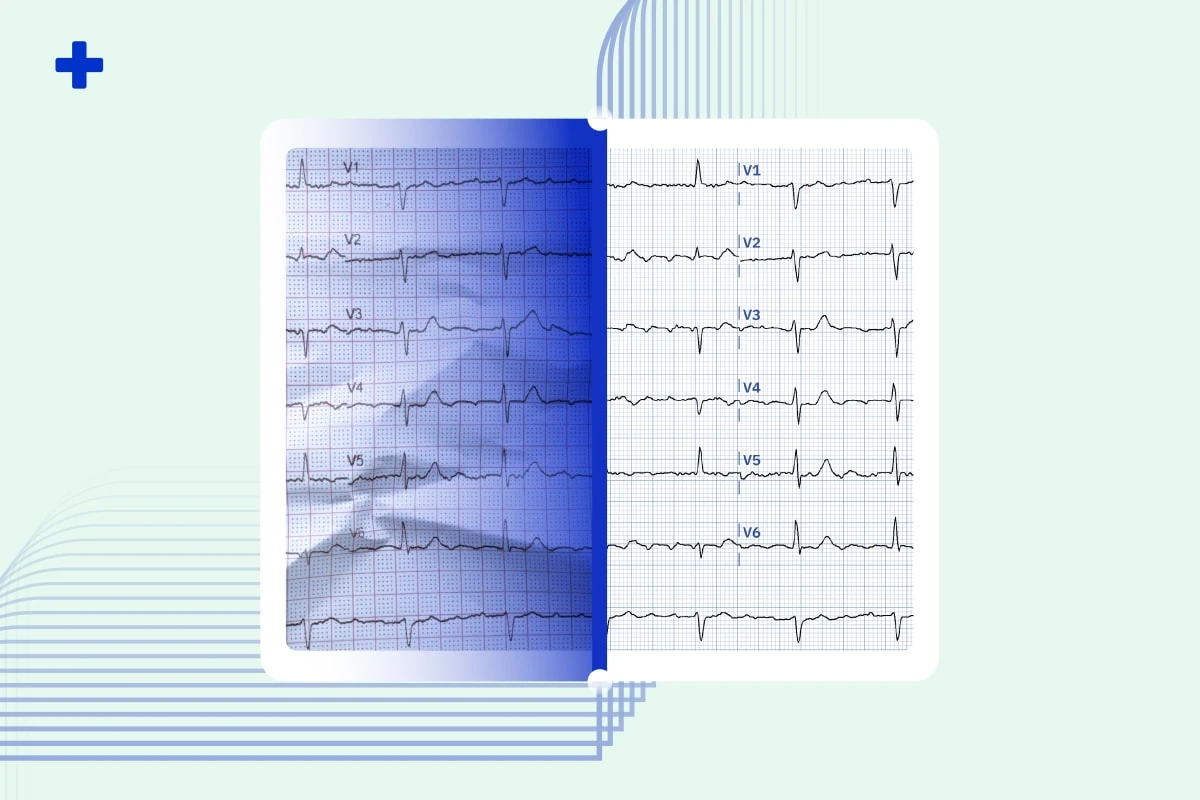
Developed a complete ECG digitization system, reconstructing ECG curves from scanned images of printed records.
An electrocardiogram (ECG or EKG) is an electronic device that measures the electrical activity of a patient’s heart via probes (electrodes) that are attached directly to the patient’s limbs and chest. All electrical impulses generated in the heart can be measured by the ECG. This type of cardiac electrical measurement is one of the most common methods cardiologists use to evaluate patients. The resulting ECG graph conveys a large amount of information about the patient’s cardiovascular health. A trained clinician can use the graph to detect issues such as cardiac rhythm disturbances (atrial fibrillation and ventricular tachycardia), inadequate coronary artery blood flow (myocardial ischemia and myocardial infarction), and electrolyte disturbances (hypokalemia and hyperkalemia).
ECG machines have been in development since the late 19th century and entered widespread use by the late 1930s. Many advances have been made since then; in the understanding of the anatomic, pathological, electrophysiological, and genetic information underlying ECG findings; and in the clinical correlations of ECG abnormalities.
Our CMO, Robert Herman, MD, discusses the future roadmap for leveraging AI in the ECG interpretation landscape on the Mayo Clinic podcast.
However, while advancements in research were made, there has not been a comprehensive updating of ECG standards and criteria since1978.
The lack of standards inevitably lead to differences in ECG record formatting. The ECG devices we find in most modern hospitals today come from a variety of manufacturers, each with their own unique ECG format — making analysis, processing, and storage across multiple formats unnecessarily difficult and in some cases even impossible. Signal graphs recorded on devices with differently configured electrode ranges may appear misleadingly similar, patient information annotations vary, the devices may use different voltages, they can be either fully digitized or still rely on paper graphs and so on.

Most ECG manufacturers produce custom-branded software for doctors to view the ECG recording on a computer. However, this software doesn’t allow the extraction of data points, which is required for data analysis on a larger scale. Vague attempts at designing digitized formats have been made by some manufacturers, but these formats mostly ended up being discontinued or deprecated. In this chaotic system most hospitals and telemedicine companies opt for the less technological but more reliable ECG storage method: printing them out and storing them in archives.
Having ECG records stored on paper in hospital archives is rather inconvenient for further research and development. We faced this issue ourselves during the development of an application for the diagnosis and management of patients with atrial fibrillation. One of the incremental modules of the application is diagnosing atrial fibrillation based on ECG recordings.
This roadblock lead to the development of a full-fledged general ECG digitization system, which reconstructs the ECG curve directly from a scanned image of a printed record. Our system supports a wide range of non-signal files, including any image file format and even PDF files, converting them all into a single unified format of labelled data points.
The implications of bringing ECG records into the digital space are substantial. Creating vast datasets of labeled ECG records will allow unimpeded use of machine learning for any analysis or classification (potentially assisting doctors in the diagnostic process). It will open the data to further valuable research. There is also great demand for such a system in the healthcare sector simply for digitizing and storing their existing archived ECG records. Our software will allow them to process these various formats and offer insightful analysis.
Share this article
About PMcardio
PMcardio is a CE-marked Class IIb AI medical device that interprets any 12-lead ECG — from a photo or a digital file — in seconds. It detects 50+ ECG findings, including occlusion myocardial infarction (OMI) — the acute coronary occlusions that conventional STEMI criteria miss in over half of cases. Its Queen of Hearts™ OMI model holds FDA Breakthrough Device Designation, and PMcardio is already used by 100,000+ clinicians who have analyzed more than 2.5 million ECGs.
About Powerful Medical
Powerful Medical is a medical-technology company using artificial intelligence to transform how cardiovascular disease — the world’s leading cause of death — is diagnosed. Founded in 2017, it built PMcardio, the world’s first AI medical device to detect acute coronary occlusion (OMI) from an ECG. Its research is published in the European Heart Journal – Digital Health and validated across 25+ clinical studies, and its Queen of Hearts™ model earned FDA Breakthrough Device Designation. To date, PMcardio has supported the detection of 120,000+ heart attacks across 150+ hospital deployments worldwide.