

Bifascicular Block Identified as STEMI-Equivalent by Queen of Hearts
This case report highlights that in the right clinical context, early recognition of bifascicular block (BFB) as a STEMI-equivalent or occlusive MI pattern is crucial, since it often signals proximal LAD occlusion, large myocardial injury, and a poor prognosis. It also offers initial real-world evidence that the AI-based Queen of Hearts app can reliably support clinical decision-making in acute coronary syndrome care.
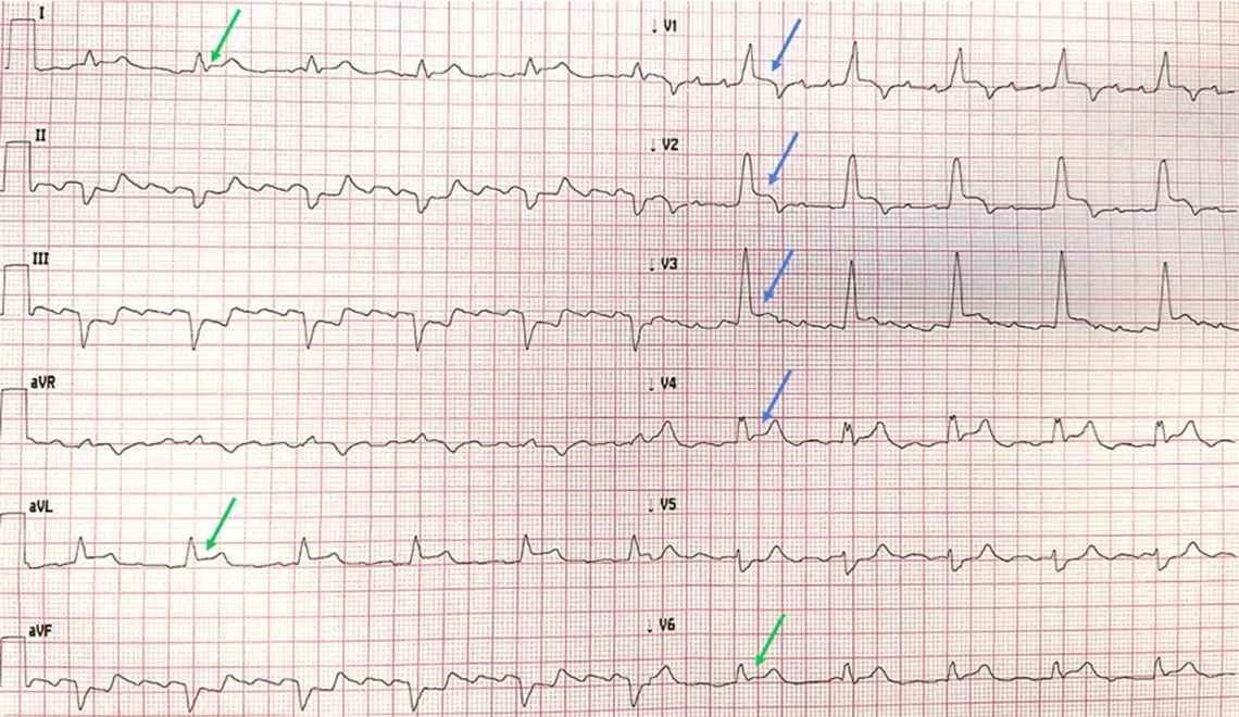
A man in his 70s presented with chest pain and hypotension suggestive of acute coronary syndrome. The ECG demonstrated bifascicular block (right bundle branch block and left anterior fascicular block) with QRS-concordant ST elevation in the anterior and lateral leads.
ECG
AI ECG Interpretation
The ECG was analyzed using the AI-based Queen of Hearts application (PMcardio). The algorithm identified the STEMI-equivalent pattern, detected atrial flutter and bifascicular block, and predicted reduced left ventricular ejection fraction. These findings supported the suspicion of an occlusive myocardial infarction requiring urgent reperfusion.
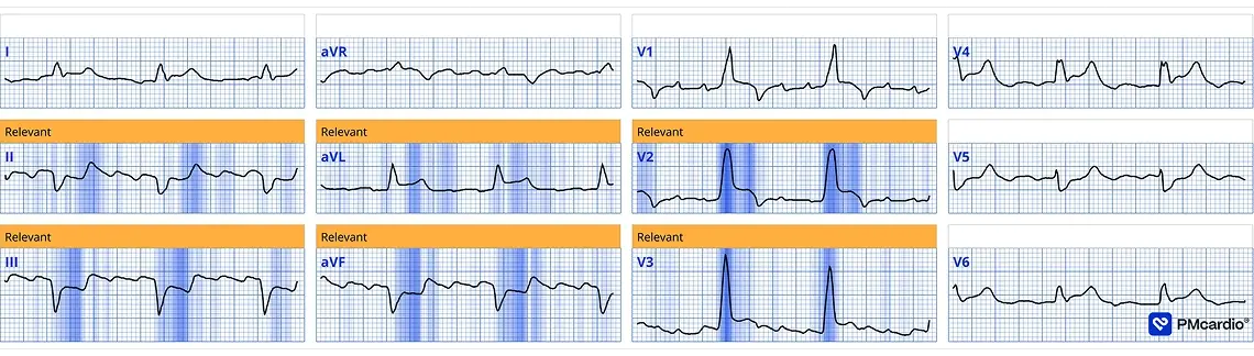
AI ECG Interpretation
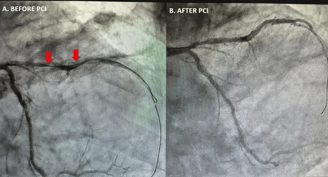
The patient was transferred for emergent coronary angiography. The procedure confirmed a near-occlusive proximal LAD lesion. Primary percutaneous coronary intervention with drug-eluting stent implantation successfully restored coronary flow.
This case illustrates how bifascicular block in the setting of ischemic symptoms can represent a STEMI-equivalent pattern associated with proximal LAD occlusion. Early recognition of this high-risk ECG pattern, supported by AI-assisted ECG interpretation, can facilitate timely diagnosis and rapid activation of reperfusion therapy.
Share this article


